General Topics

Self-assessment
The following true/false questions may be attempted before reading the tutorial. The answers are given at the end of the text.
- In the normal heart:
a. Pre-load is the same as left ventricular end-diastolic volume
b. Increase in pre-load has no effect on the force of the next contraction
c. Systemic vasoconstriction reduces afterload
d. Isovolumetric ventricular contraction ends when the aortic valve opens
e. The ejection fraction of a normal left ventricle is approximately 40% - Regarding diastole:
a. Diastole is an active, energy consuming process
b. Diastolic time remains the same with increased heart rate
c. In the normal heart, only one third of ventricular filling occurs before atrial contraction
d. Atrial contraction is a part of diastole
e. Ventricular relaxation is a part of diastole - Regarding heart failure:
a. The failing ventricle is invariably more compliant than normal
b. Central venous pressure should be kept to a minimum if diastolic failure is suspected
c. Allowing tachycardia is an effective way of maintaining cardiac output
d. Vasodilation can help restore cardiac output in the failing ventricle
e. Inotropes should never be used - Regarding the management of patients with heart failure:
a. ACE inhibitors should normally be continued throughout the perioperative period
b. Beta-blockers should be normally be continued throughout the perioperative period
c. Arrhythmias should only be treated if blood pressure is compromised
d. Spinal anaesthesia reduces perioperative risk in total knee replacement surgery
Introduction
Congestive cardiac failure (CCF) is a common and debilitating condition. It is characterised by impaired ventricular performance resulting in fatigue, exercise intolerance, an increased incidence of ventricular arrhythmias and a shortened life expectancy. The three major risk factors for the development of heart failure are age, hypertension and coronary artery disease. In the UK up to 4% of deaths are attributable to heart failure and 40% of patients will die within one year of diagnosis. Perioperatively, heart failure is associated with a substantial increase in morbidity and mortality.
This review will examine the underlying pathophysiological principles of CCF and apply those principles to the clinical management of patients.
Pathophysiology
Characteristically the chambers of the heart become enlarged, with increased wall thickness and stiffness. Underlying this is a process of fibrosis and myocellular hypertrophy. These morphological changes lead to important functional changes which affect both diastole (relaxation) and systole (contraction).
Diastole
Figure 1 shows the normal left ventricular pressure-volume relationship (LVPVR) during the cardiac cycle. Diastole can be divided into three stages:
Isovolumetric ventricular relaxation: This is demonstrated by the vertical descending part of the pressure-volume loop. It is an active process requiring ATP, in which calcium is sequestered back into the sarcoplasmic reticulum. In diastolic heart failure this process breaks down, resulting in a ventricle which fails to relax and is therefore stiff and less compliant.
Passive ventricular filling: In the normal heart, approximately 75% of ventricular filling (preload) occurs passively.
Atrial contraction: In the normal heart, atrial contraction contributes only a quarter of left ventricular end-diastolic volume (LVEDV) or preload. However, for a ventricle which is poorly compliant, due to diastolic failure, preload is much more dependent on atrial contraction.
The dotted line in figure 1 shows how the mechanics of the LVPVR change in diastolic heart failure. For any given left ventricular volume, there is a higher left ventricular pressure. This has two major consequences which need to be understood. Firstly, there will be a decreased left atrial (LA) to left ventricular (LV) pressure gradient. LV filling in diastole depends on this pressure gradient. If LA pressure drops, LVEDV (preload) diminishes rapidly, along with stroke volume and cardiac output. Hypovolaemia is therefore poorly tolerated. (A note of caution here – the situation is quite different in acute LV systolic failure, when a reduction of preload can potentially rescue the ventricle. This is explained later). Secondly, there will be a susceptibility to develop pulmonary oedema, as the increased left sided heart pressures result in increased pulmonary venous and capillary pressures. Up to a half of patients presenting with respiratory symptoms of congestive heart failure will have preserved systolic function.
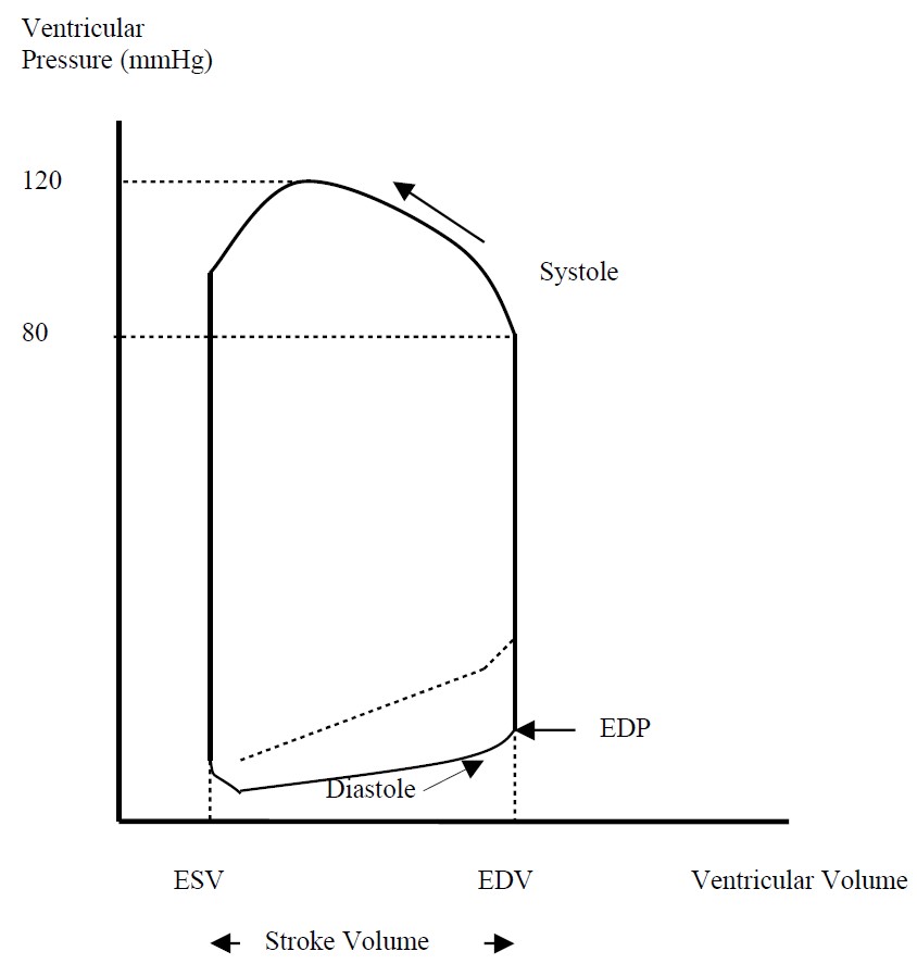
Figure 1. Left ventricular pressure-volume relationship (Dotted line indicates diastolic dysfunction)
Systole
Systole consists of two stages. The first stage consists of isovolumetric ventricular contraction, which ends with the opening of the aortic valve and is represented by the vertically ascending portion of the loop. The second stage concerns the ejection of blood into the aorta and ends with the closure of the aortic valve. These are ATP consuming, active processes involving the interaction of actin with myosin. Systolic heart failure occurs when there is inadequate force generation to eject blood normally. It can affect either ventricle but left-sided heart failure is more common. Systolic failure is likely to co-exist with diastolic failure.
Reduction in cardiac output, whether acute or chronic, induces a neuro-endocrine response. Baroreceptor activity increases efferent sympathetic nervous system (SNS) activity and circulating catecholamines increase. The SNS activates the reninangiotensin- aldosterone (RAA) system. These responses initially act to maintain cardiac output and perfusion pressure but ultimately their effects become deleterious. SNS activity increases myocardial oxygen consumption, which may not be met by supply. Along with a contribution from angiotensin II, the SNS also increases afterload and therefore myocardial work. Sodium and water retention by the kidneys may have an adverse influence on preload and exacerbate oedema. These processes are attenuated in the medical management of congestive cardiac failure by betablockers, ACE inhibitors and diuretics.
Figure 2 shows the relationship between LVEDV (or preload) and stroke volume (SV) described by Frank and Starling. The dotted line shows the effect of an increasing preload on a failing ventricle. The progressive decrease in SV as a result of overstretched, diseased cardiac muscle fibres results in a higher and higher LVEDV, amplifying the problem. This describes the situation in acute LV failure which can be managed using the vasodilatory properties of intravenous nitrates. Venodilation reduces preload with the aim of bringing the ventricle back along the curve to a point of increased SV at a decreased LVEDV. Dilation on the arterial side reduces afterload and therefore myocardial work.
The effect of increased afterload can also be understood by looking at the Frank- Starling graph. The arrow downwards from the point marked with a cross represents the immediate effect of an increase in afterload. The next contraction of the heart will yield a smaller SV as it is pumping against a higher vascular resistance. A smaller SV results in a lower ejection fraction and therefore more blood remains in the ventricle. This results in a greater LVEDV (preload) for the next contraction. The next arrow follows the mechanical path such a ventricle takes next – along the Starling curve to the right. In a good ventricle, SV is regained (at a higher LVEDP). If the ventricle is failing, however, an increased afterload can lead to rapid decompensation (further down the dotted lined curve to the right).
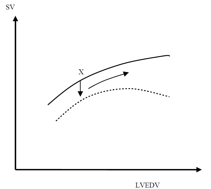
Figure 2. Frank-Starling curve (Dotted line indicates performance of failing ventricle)
Anaesthetic management of patients with heart failure
Preoperative Assessment
It is important that patients with congestive cardiac failure are identified preoperatively, especially those with evidence of current or recent decompensation. Evidence of decompensation within 6 months of surgery is associated with increased risk. There may be a history of shortness of breath and reduced exercise tolerance. Patients unable to sustain 4 ‘metabolic equivalents’ (climbing a flight of stairs) are particularly at risk from perioperative complications. Associated co-morbidity such as ischaemic heart disease, hypertension and diabetes should be sought. Examination may reveal peripheral or pulmonary oedema and a third heart sound.
Investigations
Blood tests: Anaemia and electrolyte disturbance (especially for patients taking diuretics) should be identified and treated. Other blood tests which may reveal aggravating factors include liver and thyroid function tests and glucose.
ECG: Check for arrhythmias.
CXR: Signs may include cardiomegaly, pleural effusions, prominent upper lobe veins (upper lobe diversion), engorged peripheral lymphatics and alveolar oedema.
Transthoracic echocardiogram: This is the most useful test and can provide important anatomical information as well as an assessment of function. Heart failure can be secondary to valvular disease (usually aortic stenosis or mitral regurgitation). Patients with an ejection fraction of less than 40% are considered to have systolic failure and those with an ejection fraction of less than 30% have severe disease.
Cardiac catheterisation: May be performed if significant coronary or valvular heart disease is suspected as the cause of heart failure.
Optimisation of treatment
Medical therapy should be optimised to minimise symptoms of left ventricular failure and maximise functional capacity. Along with diuretics, many patients with CCF will be taking a beta-blocker and an ACE inhibitor (or Angiotensin II receptor antagonist). These drugs reduce myocardial work by controlling heart rate and reducing afterload respectively. Many anaesthetists stop ACE inhibitors on the day of surgery due to problems with blood pressure lability, but all other anti-failure therapy should be continued in the perioperative period. There is some evidence that perioperative betablockade reduces morbidity and mortality in patients at high risk of cardiac complications. Symptomatic arrhythmias should be treated, and attempts made to control heart rate to around 80 beats per minute. Atrial fibrillation is poorly tolerated.
Elective surgery should be postponed until medical management has been optimised and risk assessed thoroughly against benefit. High risk patients undergoing elective or emergency surgery may benefit from preoperative optimisation in ICU or HDU.
Intraoperative Management
Patients undergoing minor peripheral procedures should be offered local or regional anaesthesia where possible. For more major surgery there is no evidence of the benefits of general versus regional anaesthesia. However, whichever technique is used, having understood the pathophysiology of cardiac failure, there are clear haemodynamic goals for the perioperative period. These are aimed at preserving cardiac output and minimising myocardial work.
- Preserving cardiac output: There are three factors that influence cardiac output. These are preload, afterload and contractility. The poorly compliant ventricle must be given the opportunity to fill in diastole. This will require a higher than usual central venous pressure, the avoidance of tachycardia (which reduces the duration of diastole) and in particular the aggressive treatment of arrhythmias. As discussed earlier, in the failing ventricle LVEDV is heavily reliant on atrial contraction. If this ‘atrial kick’ is lost, as occurs in atrial fibrillation for example, preload will be reduced with consequent decrease in cardiac output. Increases in afterload, especially acutely, can cause a dramatic reduction in cardiac output and should be avoided. Finally, contractility must be maintained. Patients with cardiac failure may rely on increased sympathetic tone to maintain cardiac output, and are therefore susceptible to circulatory collapse if this is lost after induction of anaesthesia. Agents such as ephedrine should be readily available although great care should be taken with vasopressors. Inotropes such as dobutamine or phosphodiesterase inhibitors may be required for patients who decompensate in the perioperative period.
- Minimising myocardial work: Tachycardia increases myocardial oxygen demand and therefore should be avoided. Consideration should be given to factors which may precipitate tachycardia, including intubation, surgical stimulus, hypovolaemia, anaemia, hypoxia, hypercapnia, post-operative pain, nausea and vomiting. Opioids such as alfentanil attenuate the response to intubation. Effective analgesia is important and use of regional techniques should be considered. Epidural infusions obtund the stress response to surgery and can provide effective post-operative analgesia. The haemodynamic effects are potentially favourable. Reducing afterload by vasodilation can greatly reduce myocardial work. However, it is important not to compromise blood flow to circulations which have pressure-dependent autoregulation (cerebral, renal and coronary). In particular, diastolic pressure must be maintained, as the left ventricle is perfused during diastole and many patients with cardiac failure will also have coronary artery disease.
With these physiological principles in mind, invasive monitoring, including cardiac output measurement, should be considered for all major surgery.
Postoperative care
All patients should receive supplemental oxygen in the postoperative period. Careful fluid balance is also required. Patients with heart failure are susceptible to renal failure due to reduced glomerular filtration rates. If urine output falls, adequate volume status, perfusion pressure and cardiac output should be ensured before diuretics are used. NSAIDs should be avoided or used with great caution to avoid further renal insult. ACE inhibitors should be reintroduced as soon as possible. If omitted for more than 3 days they should be restarted at a lower dose to avoid hypotension. There should be a low threshold for admission to ICU or HDU.
Conclusion
CCF is a major cause of morbidity and mortality worldwide and is increasing in incidence. More patients with the condition are presenting for surgery in both elective and emergency settings. Good understanding of the underlying physiological principles is necessary to manage these patients appropriately in the perioperative period and minimise their risk of complications.
MCQ Answers
- TFFTF
- TFFTT
- FFFTF
- FTFF
References
- Magner J, Royston D. Heart failure. Br J Anaesth 2004, 93: 74-85
- Pirracchio R et al. Diastolic heart failure in anaesthesia and critical care. Br J Anaesth 2007, 98: 707-21
- Pinnock C, Lin T, Smith T. Fundamentals of Anaesthesia. London: GMM Ltd, 2003
- Allman K, Wilson I. Oxford Handbook of Anaesthesia. Oxford: OUP, 2006 ATOTW
This work by WFSA is licensed under a Creative Commons Attribution-NonCommercial-NoDerivitives 4.0 International License. To view this license, visit https://creativecommons.org/licenses/by-nc-nd/4.0/