
QUESTIONS
Before continuing, try to answer the following questions. Each question has only ONE answer. The answers can be found at the end of the article, together with an explanation.
- The following statements pertain to asthma (true or false):
- the presence of wheezing is diagnostic
- one distinctive feature is airway hyper-responsiveness
- inhaled adrenaline is an effective therapy
- it can be triggered by exercise alone
- no confirmatory diagnostic test exists
- Mild intermittent asthma is characterised by the following (true or false):
- daytime symptoms ≤ twice per week
- nighttime symptoms ≤ twice per month
- PEFR/FEV1% ≥ 80% predicted
- PEFR variability ≤ 50% predictedDuring general endotracheal anaesthesia of a patient with asthma, signs of intraoperative
- During general endotracheal anaesthesia of a patient with asthma, signs of intraoperative bronchoconstriction include the following (true or false):
- upsloping of the end-tidal CO2 waveform
- hypoxaemia
- decreased peak airway pressure
- wheezing
INTRODUCTION
Asthma is a leading cause of morbidity in children throughout the world. In most hospitals in urban areas it the most common reason for hospital admission. The prevalence among children in Western countries is between 2 and 10%.1 Asthma is a lung disease that is characterized by three distinct features:
- airway obstruction
- airway inflammation
- airway hyper-responsiveness
Asthma manifests as episodes of recurrent wheezing, chest ‘tightness’, dyspnoea and dry cough. Typically episodes result in variable obstruction of airflow that may resolve either spontaneously or with treatment. Asthma has no radiologic, histologic or confirmatory blood test. It is characterized by reversible symptoms, findings on examination and pulmonary function tests. While the characteristic symptom of asthma is wheezing (usually expiratory), it is important to realize that wheezing is produced simply by airflow passing through a sufficiently narrow airway. Wheezing can occur during different portions of the respiratory cycle depending on the site of the airway obstruction and its cause. These causes can be divided into those that result most typically in inspiratory wheezing (i.e. causes of stenosis, such as tumors and foreign bodies; also hypersensitivity pneumonitis) and expiratory wheezing (i.e. asthma, bronchiolitis and COPD), with their differentiation completed on the basis of history, physical examination and pulmonary function tests.2
An acute exacerbation of asthma can quickly progress to severe respiratory distress with signs of tracheal, intercostal and subcostal retractions, use of accessory muscles of breathing, and a prolonged expiratory phase. This may ultimately lead to respiratory failure defined by hypoxaemia/hypercarbia. It is also important to note that as an acute asthma attack worsens and the child fatigues wheezing may become diminished or be completely absent. Many children with chronic asthma also have chronic inflammatory changes that may be associated with permanent alterations in their airway structure. These patients may not be responsive to commonly used treatment modalities.
EPIDEMIOLOGY
The median age of onset for asthma is 4 years old with more than 20% of children developing symptoms within the first year of life.1 Asthma most likely results from an interaction between both an inherited modifier of inflammation and environmental influences. A number of risk factors have been identified but the best researched include gender, atopy, allergens, infections, obesity, tobacco smoke, and perinatal factors.
Childhood asthma tends to occur predominantly in males until puberty. After age 20 the prevalence equals between the sexes. The association between asthma and other atopic diseases has been well researched. An “atopic march” has been identified which starts as atopic dermatitis as an infant, followed by allergic rhinitis and asthma as an older child or adolescent. Recent research has shown that indoor allergens play a significant role in the development of asthma. The dust mite, alternaria mould, cockroach allergens, as well as cat and dog allergens have all been implicated. Viral and bacterial infections are well known triggers of asthma exacerbations but their causal relationship remains unproven. Several large studies have suggested that patients with an elevated BMI or that are actively smoking are at increased risk of developing asthma. Lastly, perinatal risk factors have been studied extensively but have so far found few strong correlations – most likely due to the inherent difficulty in controlling for confounders between study groups.1
PATHOPHYSIOLOGY
The chronic airway obstruction seen in asthmatic patients is caused by inflammation and hypertrophied bronchial smooth muscle that leads to hyperinflation and air trapping. This subsequently results in a decrease in lung compliance and an increase in the patient’s work of breathing. Chronic airway obstruction leads to a ventilation / perfusion mismatch and dead space ventilation which are clinically evident as hypoxaemia and further increase the work of breathing. There may be hypocarbia initially, but hypercarbia may develop with increasing obstruction and fatigue. Hypercarbia is typically a sign of impending respiratory failure. Prolonged respiratory acidosis from hypercarbia may lead to pulmonary hypertension, which has implications on right heart function.1
PREOPERATIVE MANAGEMENT
ASSESSMENT
Important questions when evaluating a child with asthma include the following: frequency of symptoms, amount and purulence of sputum, use and effectiveness of medications, asthma triggers, activity level, previous history of surgery and anaesthesia, recent upper respiratory infections, hospitalizations and emergency department attendance.3 Hospitalizations should be investigated more thoroughly to determine if any ICU admissions or intubations were required. A complete and comprehensive examination should focus on wheezing, cough, use of accessory muscles, respiratory rate, cyanosis, and changes in mental status. Oxygen saturation (SpO2) is useful to have preoperatively. Laboratory tests are usually not required, especially for mild asthma, unless an acute infection is present. A baseline electrocardiogram and chest x-ray may be helpful (if available) for a severely asthmatic child who may have chronic airway obstruction with resultant pulmonary hypertension and right heart strain.3
Parents should be instructed to administer all asthma medications including on the morning of surgery to ensure optimal treatment prior to surgery. Ideally children with asthma who are scheduled for elective surgery should not have wheezing on the morning of surgery.
TREATMENT
An understanding of the patient’s asthma severity is critical to administering the appropriate preoperative treatment. The table below defines each severity classification based on frequency of daytime symptoms, frequency of night time symptoms, ratio of Peak Expiratory Flow Rate (PEFR) to Forced Expiratory Volume over 1 second (FEV1) and the PEFR variability.
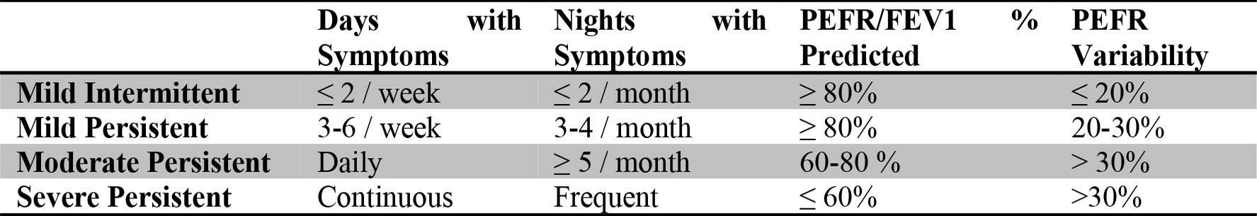
Table 1: Severity Classification of Chronic Asthma
Forced expiratory volume over 1 second (FEV1), Peak Expiratory Flow Rate (PEFR), adapted from Oski’s Pediatrics1
Preoperative treatment for mild intermittent or mild persistent asthma involves administering a nebulized β2-adrenergic agonist, such as albuterol (salbutamol), 1 to 2 hours prior to surgery.
Preoperative treatment for moderate persistent asthma involves additional optimization with any inhaled anti-inflammatory agent (suppressing the inflammatory cytokines that cause airway hyperreactivity) and by consistent use of nebulized β2 agonists 1 week prior to surgery.
Preoperative treatment for severe persistent asthma involves a visit to their primary care physician prior to surgery in order to optimize treatment. Some children benefit from short-term oral corticosteroid therapy, such as prednisone for 3 to 5 days or oral dexamethasone for 2 days prior to surgery.
The aforementioned preoperative treatments have all been shown to be both effective and safe with a low incidence of side effects.1 However, determining the severity of the patient’s asthma is not sufficient. It is also imperative to determine the patient’s control of their disease. It is possible that a patient with severe asthma may be well controlled, whereas a patient with mild asthma may be very poorly controlled. The potential for perioperative complications is present with both of these alternate scenarios.
Midazolam is a safe anxiolytic in this patient population and does not alter bronchial tone.
INTRAOPERATIVE MANAGEMENT
CHOICE OF INDUCTION AGENT
A thorough understanding of the physiological effects of each anaesthetic drug and its interaction with an asthmatic patient is crucial. Below is a description of this as it pertains to each drug and/or drug class.
Propofol inhibits bronchoconstriction and increases central airway dilation by directly relaxing the airway smooth muscle. These actions decrease the possibility of bronchospasm during induction. However, this agent may not be suitable for haemodynamically unstable patients. This agent is considered safe for patients with asthma.
Ketamine has sympathomimetic bronchodilatory properties, with a direct action on bronchial smooth muscle that causes a relaxation of bronchiolar musculature. These actions decrease the possibility of bronchospasm with induction and therefore ketamine has been proposed as a good choice for patients with severe persistent asthma.4 However, ketamine increases bronchial secretions and must be given simultaneously with an anticholinergic drug such as glycopyrrolate or atropine.
Thiopental use has been associated with bronchospasm and is therefore not a good first choice for induction of anaesthesia.
Lidocaine (lignocaine), when given intravenously, significantly increases the histamine threshold and blocks the cough reflex. It may be given to decrease the airway responses associated with endotracheal intubation.
Halothane (plus enflurane/isoflurane) are potent bronchodilators that act via β-adrenergic receptor stimulation. They have been shown to decrease airway responsiveness and help ease histamineinduced bronchospasm. Many studies have shown their effectiveness in the treatment of status asthmaticus.5 Sevoflurane has controversial effects in asthmatics with some studies showing an increase in airway resistance and others showing no change.4 Desflurane is a very pungent agent that is irritable to the airway and has been shown to increase secretions, coughing, and laryngospasm. Halothane and sevoflurane remain good choices for inhalational induction.
Morphine causes histamine release which may result in bronchospasm. Fentanyl, if rapidly administered in large doses, can result in chest rigidity which could be mistaken for bronchospasm
The neuromuscular blockers that bind and stimulate M2 muscarinic receptors more than M3 muscarinic receptors, such as gallamine, pipecuronium and rapacuronium, cause bronchoconstriction. The neuromuscular blockers that stimulate the M2 and M3 muscarinic receptors evenly, such as vecuronium, rocuronium, cisatracurium and pancuronium, do not cause bronchoconstriction.4 Atracurium and mivacurium dose-dependently release histamine and thus trigger bronchoconstriction.
INTUBATION TECHNIQUE
Airway manipulation particularly in a light plane of anaesthesia is a potent stimulus of bronchospasm, laryngospasm, coughing and breath-holding. Use of an inhaled β2-agonist before intubation will help reduce bronchospasm. In general, avoidance of any stimulation of the airway seems sensible. For short cases a face mask may be sufficient. Whenever possible avoidance of tracheal intubation is preferred and a laryngeal mask airway (LMA) should be used since it may be less stimulating than an endotracheal tube. However, in cases that require airway protection (for instance patients with severe gastro-oesophageal reflux disease), a LMA should not be used. If endotracheal intubation is necessary, ensuring a deep level of anaesthesia prior to airway instrumentation reduces the risk of bronchoconstriction.3 A tracheal tube that is too long and touches the carina is a potent cause of bronchospasm; the length of the tracheal tube should therefore be assessed carefully after intubation.
MAINTENANCE
Maintenance of anaesthesia with inhalational agents such as halothane helps to dilate bronchioles and prevent bronchospasm. It is important to have a method to deliver a bronchodilator intraoperatively after the airway is secured. A simple technique is to remove the plunger from a 50ml syringe and place a bronchodilator metered dose inhaler (MDI) into the barrel. The plunger is then replaced and when depressed actuates the MDI to spray through the Luer connector of the syringe. This can then easily be incorporated into the anaesthesia circuit. Specific adapters and nebulising circuits have been made for this purpose.3
One must be vigilant for the signs of intraoperative bronchospasm in order to be able to treat it. Intraoperative bronchospasm presents as a prolonged expiratory phase/wheeze with increased airway pressure, hypoxia and upsloping in the end-tidal CO2 waveform and may result in difficulty with ventilation. Other causes of airway obstruction (e.g. equipment malfunction) and wheeze (e.g. pulmonary aspiration) must be ruled out prior to initiation of treatment with an inhaled β2-agonist such as albuterol (salbutamol). The first intervention for intraoperative bronchospasm is to remove the causative stimulus while increasing the FiO2, increasing expiratory time and deepening the anaesthetic. The wave-form of the end-tidal CO2 waveform is an early indicator of the effectiveness of these interventions.6
EXTUBATION
For patients undergoing elective surgery and where no other contraindications exist, deep extubation is a reasonable option. For successful deep extubation the depth of anaesthesia should be sufficient to prevent laryngo-/bronchospasm when the endotracheal tube is removed. For patients undergoing emergency surgery, with a potentially full stomach, awake extubation is a more sensible option. An awake extubation occurs after upper airway reflexes have returned and the patient can therefore protect their airway. Sufficient amounts of opioid and redosing of inhaled β2-agonists may also be useful prior to extubation in these circumstances to decrease the reflex bronchoconstriction that may occur. It may be preferable to avoid reversal of muscle relaxants at the completion of surgery. Both neostigmine and physiostigmine cause increased secretions and bronchial hyperreactivity. If possible, administer muscle relaxants at times and doses that enable them to wear off appropriately without the need for reversal agents.
POSTOPERATIVE MANAGEMENT
ANALGESIA
When possible and where indicated, postoperative regional analgesia is preferable. A functioning epidural will block afferent pathways mediating pain from the abdominal viscera/incision and therefore help to maintain the patient’s respiratory muscle function. This leads to more adequate tidal volume and vital capacity and helps to preserve diaphragm function.7 A regional-only anaesthetic technique also eliminates the need for airway manipulation, thus preventing any potential airway irritation associated with it. Avoidance of both meperidine (pethidine) and morphine is preferable, given their ability to release histamine which can result in bronchospasm. Fentanyl is the best opioid alternative. Ketamine infusion has also been used in the postoperative period since it is capable of providing analgesia with the direct advantage of also preventing bronchospam.8
MONITORING
Standard postoperative monitors are indicated while waiting for the patient to regain normal airway/breathing function. The head up position is preferred to help clear secretions and prevent atelectasis. The early control of sputum and respiratory symptoms helps to prevent postoperative complications.
CONCLUSION
Remembering four key points in the perioperative management of asthma can result in decreased morbidity and mortality:
- A thorough understanding of asthma . This is necessary to select the anaesthetic agents and approaches that will optimize the patient’s pulmonary function
- Proper preoperative management . One of the most effective ways to manage perioperative asthma is prevention and this starts with proper preoperative assessment and planning. Preoperative evaluation is critical to determine if bronchodilators and/or steroids are needed. Effort aimed at improving the quality of preoperative care results in well-controlled, stabilized children coming to surgery
- Intraoperative vigilance
- Prompt and early recognition of asthma exacerbations
ANSWERS TO QUESTIONS
- Concerning asthma:
- FALSE. The presence of wheezing is NOT diagnostic. Wheezing is present in numerous disease processes including bronchiolitis, cystic fibrosis, recurrent pulmonary aspiration, mediastinal masses, tracheomalacia, brochomalacia, tracheal web, tracheal stenosis, and bronchial stenosis to name a few.
- TRUE. Asthma has three distinct features: 1) airway obstruction, 2) airway inflammation, and 3) airway hyper-responsiveness.
- TRUE. Inhaled epinephrine is an effective, although not first line, therapy.
- TRUE. Asthma can be triggered by exercise alone.
- TRUE. No confirmatory diagnostic test exists for asthma.
- Concerning mild intermittent asthma:
- TRUE. Daytime symptoms occur ≤ twice per week.
- TRUE. Nighttime symptoms occur ≤ twice per month.
- TRUE. PEFR/FEV1% is ≥ 80% of the predicted value.
- FALSE. PEFR variability must be ≤ 20% (rather than <50%) of the predicted value.
- During general endotracheal anesthesia of a patient with asthma, signs of intraoperative brochoconstriction include the following:
- TRUE. An upsloping of the end-tidal CO2 waveform may occur.
- TRUE. Hypoxemia may occur.
- FALSE. Increased (rather than decreased) peak airway pressure may occur.
- TRUE. Intraoperative wheezing may occur.
REFERENCES and FURTHER READING
- McMillan J, Feigin, R, DeAngelis, C, Jones M, Oski’s Pediatrics, 4th Edition, 2006, pp. 2405- 10.
- Fanta C, Fletcher S, An overview of asthma management, UpToDate, September 30, 2009.
- Cote C, Lerman J, Todres I, A Practice of Anesthesia for Infants and Children, 2009, pp. 229- 33, 776-78.
- Burburan S, Xisto D, Rocco P, Anaesthetic Management in Asthma, Minerva Anestesiol, 2007; Vol 73, pp. 357-65.
- Restrepo R, Pettignano R, DeMeuse P, Halothane, an Effective Infrequently Used Drug, in the Treatment of Pediatric Status Asthmaticus , J Asthma, 2005; Vol. 42 (8), pp. 649-51.
- Tirumalasetty J, Grammer LC, Asthma, surgery, and general anesthesia: a review, J Asthma, 2006; Vol. 43 (4), pp. 251-54.
- Yamakage M, Iwasaki S, Namiki A, Guideline-oriented perioperative management of patients with bronchial asthma and chronic obstructive pulmonary disease , J Anesth, 2008; Vol. 22 (4), pp. 412-28.
- Jahangir S, Islam F, Aziz L, Ketamine Infusion for Postoperative Analgesia in Asthmatics: A Comparison with Intermittent Meperidine, Anesth Analg, 1993; Vol.76, pp. 45-49.
This work by WFSA is licensed under a Creative Commons Attribution-NonCommercial-NoDerivitives 4.0 International License. To view this license, visit https://creativecommons.org/licenses/by-nc-nd/4.0/