Basic Sciences

QUESTIONS
Before continuing, try to select the correct answers from the following statements. The answers can be found at the end of the article, together with an explanation.
- The normal adult kidneys:
- Lie at the level of T12 to L2 within the peritoneum
- Are approximately 9cm long
- Have an outer cortex and an inner medulla
- The left kidney has a longer renal artery
- Regarding nephrons in the kidney:
- Two distinct types are identifiable
- All have their glomeruli in the medulla
- The juxtaglomerular apparatus is part of the proximal tubule
- The collecting ducts of all nephrons pass through the medulla
- Renal blood flow:
- Is equivalent to 400ml/min/100g of tissue
- Is very high due to the kidneys high metabolic rate
- Is less per unit weight that the brain
- Is equally distributed between cortex and medulla
- Glomerular filtration:
- Occurs at a rate of 125ml/min
- Is mainly controlled by the capillary endothelium
- Results in a filtrate with the same osmolality as plasma
- Favours filtration of negatively charged molecules
INTRODUCTION
It is easy to think of the kidneys as simply excretory organs that produce urine to remove waste products from the body. In fact, they are much more complex, performing many functions which have wide ranging physiological effects. The main functions of the kidney are:
To regulate:
- Extracellular fluid volume
- Extracellular fluid electrolyte composition
- Total body water volume
- The body’s acid-base balance
- Arterial blood pressure
To produce:
- The active form of vitamin D (1, 25 dihydroxycholecalciferol)
- Renin
- Erythropoietin
- Glucose
To excrete:
- Endogenous waste products; for example, urea, creatinine, uric acid, and bilirubin
- Exogenous waste products; for example, drugs and drug metabolites
This tutorial will describe the important anatomical features and physiological processes which allow the kidneys to produce an ultrafiltrate of plasma that ultimately goes on to become urine. The details of how the kidneys alter the composition of this ultrafiltrate to regulate body fluid composition and volume will be considered in a follow-up tutorial.
GROSS ANATOMY OF THE KIDNEY
There are normally two separate kidneys each with its own fibrous capsule. They are located in a retroperitoneal location in the upper abdomen, one in each paravertebral gutter adjacent to T12 to L3. They are approximately 12cm long and weigh 150 grams each. The right kidney is slightly lower than the left due to the presence of the liver in the right upper abdomen. The upper part (upper pole) of each kidney is protected posteriorly by the 11th and 12th ribs.
The kidney has two distinct regions (see figure 1); a cortex around the outer edge, and an inner medulla. The medulla is composed of numerous renal pyramids. At the innermost ends of the pyramids are calyces which receive urine, which then drain to the renal pelvis and the ureter.
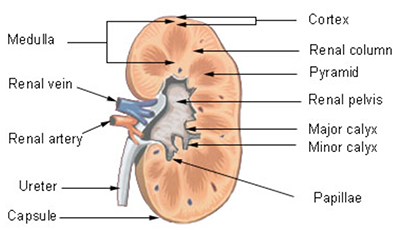
Figure 1. Diagram showing renal cortex and medulla (From Wikimedia commons: http://training.seer.cancer.gov/module_anatomy/unit11_2_uri_comp1_kidney.html)
The basic functional unit of the kidney is the nephron (see figure 2). Each kidney contains approximately 1 – 1.5 million nephrons. Each nephron is basically a folded up tube; situated proximally is a complex capillary network and capsule where plasma is filtered (the glomerulus and Bowman’s capsule), which produces the glomerular filtrate, and situated distally are the collecting ducts from which urine drains. Between Bowman’s capsule and the collecting duct is the proximal convoluted tubule (PCT), the loop of Henle and the distal tubule, each of which serve specific functions. The nephrons are all orientated such that the glomerulus and Bowman’s capsule lie in the cortex with their loop of Henle and collecting duct pointing towards and entering the medulla.
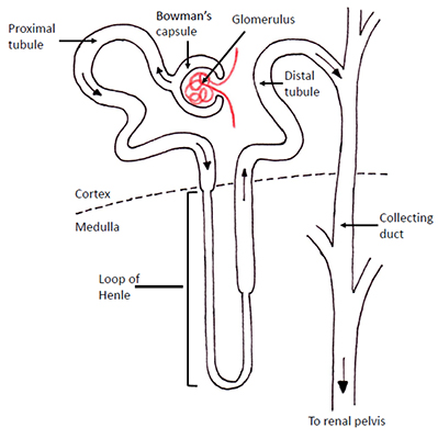
Figure 2. Configuration of a typical nephron with each distinct region labelled
ANATOMY OF A NEPHRON
Glomerulus and Bowman’s capsule
Every nephron has a glomerulus, and all glomeruli lie within the cortex of the kidney. The glomerulus is an arrangement of specialised capillaries that have an afferent arteriole at one end and an efferent arteriole at the other end. It produces an ultrafiltrate of plasma which enters the nephron tubule lumen at the Bowman’s capsule (the process of glomerular filtration is described in detail later).
Proximal Tubule
This is a continuation of the nephron tubule from the Bowman’s capsule and is divided into two parts; the convoluted (twisted or folded) proximal tubule (pars convoluta) and a later straight part (pars recta) before becoming the descending limb of the loop of Henle. The main role of the proximal tubule is reabsorption of electrolytes and water lost from the plasma through filtration at the glomerulus. It also has a role to play in secretion of substances into the tubule lumen (e.g. drugs) as well as in the regulation of in acid-base balance.
Loop of Henle
The loop of Henle descends from the renal cortex (hence descending limb) into the medulla, before doing a tight ‘U-turn’ and ascending back towards the cortex (hence ascending limb). The ascending limb comprises two distinct sections; a thin-walled section and a thick walled section (see figure 2).
Nephrons with glomeruli on the very outer edge of the cortex have short loops of Henle which only enter the outer medulla. Nephrons with glomeruli located near the corticomedullary junction have very long loops of Henle that descend deep into the medulla to the tip of the renal pyramids. These two distinct types of nephrons are called ‘cortical’ and ‘juxtamedullary’ respectively (see figure 3). The importance of these two different types of nephron will become apparent when the function of the loop of Henle is discussed in detail later.
- Cortical – account for 85% of the total number of nephrons, have short loops of Henle
- Juxtamedullary – account for 15% of the total number of nephrons, have long loops of Henle
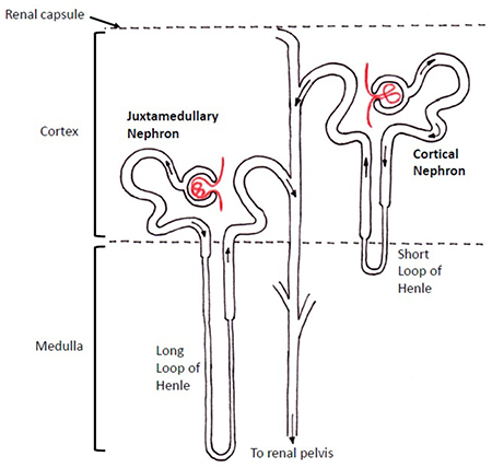
Figure 3. Diagram showing the difference between ‘cortical’ and ‘juxtamedullary’ nephrons
Juxtaglomerular apparatus
The final part of the ascending limb of the loop of Henle is located in the cortex of the kidney immediately adjacent to the afferent and efferent arterioles of its own glomerulus. This region contains the juxtaglomerular apparatus which consists of:
- Macula densa – specialised cells in the wall of the tubule at this point that are capable of sensing and responding to the composition of tubular fluid
- Afferent arteriole granular cells – specialised cells in the wall of the afferent arterioles that secrete renin
Collecting ducts
The collecting duct starts after the termination of the ascending limb of the loop of Henle in the cortex of the kidney. The collecting ducts of all nephrons pass through the renal medulla to drain the urine produced by the nephron into the calyces.
RENAL CIRCULATION
Each kidney receives its blood via the renal artery, a direct branch of the abdominal aorta (usually a single vessel but in around a quarter of individuals there are two renal arteries on each side). Venous drainage is usually via a single renal vein into the inferior vena cava (IVC). These vessels, (along with the ureter) enter the kidney via an indentation in its medial surface called the hilum. Due to the location of each kidney relative to the aorta and the IVC, the right kidney has a longer renal artery, whilst the left kidney has a longer renal vein.
Once the renal artery has entered the hilum of the kidney it divides into numerous interlobar arteries which radiate out towards the cortex (see figure 4). The interlobar arteries divide into arcuate arteries which arc around, following the line of the corticomedullary junction. The arcuate arteries give rise to several interlobular arteries which extend outwards towards the outer edge of the cortex (see figure 4). The afferent arterioles arise from the interlobular arteries, which supply blood to the glomerular capillaries. The glomerular capillaries are followed by the efferent arterioles and then the peritubular capillaries. There is a careful arrangement so that each set of glomerular capillaries and peritubular capillaries are associated with the same nephron.
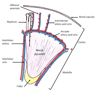
Figure 4. Diagram showing the arrangement of the intra-renal vessels
The renal circulation is unique in having a capillary bed (glomerular capillaries) with arterioles at both ends. The tone of both the afferent and efferent arterioles can be varied to influence blood flow and pressure within the glomerulus (see figure 9 a-c).
The venous system follows a similar pattern in reverse; blood flows from the peritubular capillaries into interlobular veins, arcuate veins, interlobar veins and then the renal vein.
The kidneys receive both somatic (sensory) innervation and sympathetic nervous system innervation via a ‘renal plexus’ of nerves located around each renal artery. The sensory nerves enter the spinal cord at the level of T10 or T11.
RENAL BLOOD FLOW (RBF)
The kidneys receive a total blood flow of approximately 1000mls per minute (20% of the cardiac output). This equates to 300 – 400mls per minute per 100g of tissue which is approximately six times that of the brain and five times that of the heart, weight for weight.
The blood flow is not evenly distributed throughout the kidney and is not related to the level of metabolic activity. The cortex receives 90% of blood flow, which is the least metabolically active, while only 10% goes to the more metabolically active medulla. Consequently the cortex has “luxury perfusion” with blood flow equating to ten times what is needed for oxygen delivery, whilst flow to the inner medulla is barely adequate to meet the oxygen demands.
- Cortex blood flow – 500ml/min/100g
- Outer medulla blood flow – 100ml/min/100g
- Inner medulla blood flow – 20ml/min/100g
The reason for such a seemingly excessive blood flow, particularly to the cortex, is that this is what is required to drive filtration of plasma at the glomerulus at an adequate rate i.e. provide an adequate glomerular filtration rate (GFR).
THE GLOMERULUS AND ITS FUNCTION
The glomerulus essentially acts as a filter, producing an ultrafiltrate of the plasma from the glomerular capillaries that enters the Bowman’s space. Filtration is the bulk flow of solvent through a filter carrying with it all the solutes small enough to pass through the filter. The term ‘ultrafiltration’ simply means that this process is happening on a molecular level.
The structure of the filter
The glomerular filter is made of three distinct layers, each fulfilling separate functions (see figure 5):
- The glomerular capillary endothelium:
- A highly specialised capillary endothelium with fenestrations (windows) to minimise the filter thickness;
- This layer prevents cellular components of blood coming into contact with the basement membrane.
- The glomerular basement membrane:
- Made of connective tissue, it is negatively charged;
- This is the layer that actually acts as the filter.
- Bowman’s epithelial cells (podocytes):
- Epithelial cells with multiple projections (foot processes) which interlink with each other whilst still keeping a small gap between them creating a large surface area;
- Act to maintain the basement membrane, and has phagocytic functions.
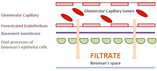
Figure 5. Diagram showing the different layers across which the filtrate must pass within the glomerulus
The degree to which solutes are filtered is dependent on two physical properties (see figure 6):
- Molecular weight
- Less than 7000 Daltons – molecules will be freely filtered
- Greater than 70 000 Daltons – molecules are essentially not filtered at all
- In between 7000 and 70 000 Daltons – the percentage of a given molecule that is filtered decreases with increasing weight
- Electrical charge
- For any given molecular weight between 7000 and 70 000 Daltons a lower percentage of negatively charged molecules will be filtered
- This is due to the basement membrane having a negative charge and therefore repelling negatively charged molecules
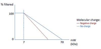
Figure 6. Graph showing the influence of molecular weight and charge on degree of filtration at the glomerulus
The cellular components of blood are prevented from being filtered by the capillary endothelium, and the proteins present within plasma are almost entirely prevented from being filtered by their size and their negative charge.
A proportion of the plasma passing through the glomerulus freely passes through the filter taking with it very small molecules such as dissolved electrolytes (e.g. sodium, potassium, bicarbonate) and other solutes (e.g. glucose, urea). Hence, the ultrafiltrate fluid in the Bowman’s space has the same concentration of electrolytes, glucose and urea as in plasma. It also means that the plasma that is not filtered, but which remains within the vascular system is essentially unaltered in terms of its osmolality and electrolyte composition, but does have a slightly higher haematocrit and protein concentration.
What determines GFR?
The rate at which filtration occurs depends upon:
- The surface area of the filter;
- The thickness or permeability of the filter;
- Magnitude of any forces favouring filtration;
- Magnitude of any forces opposing filtration.
For the glomerulus, the surface area and the permeability of the filter can be combined into a numerical constant called the “coefficient of filtration” (represented as ‘Kf’). The forces favouring or opposing filtration at the glomerulus follow the same principles as for any capillary; capillary and interstitial hydrostatic pressures (Pcap and Pi) and capillary and interstitial oncotic pressures (Πcap and Πi). When considering the glomerulus, the interstitial space refers to Bowman’s space (and the forces acting are represented by the symbols PBS and ΠBS accordingly). Fluid will tend to move from an area of higher to lower hydrostatic pressure and from an area of lower to higher oncotic pressure. This can all be summarised in words and in symbols as:
Glomerular Filtration Rate (GFR) = Kf {(forces favouring filtration) – (forces opposing filtration)}
GFR = Kf {(Pcap + ΠBS) – (PBS + Πcap)}
The forces favouring and opposing filtration will differ in magnitude from the proximal to the distal end of the capillary. For this reason the forces involved at each end of the capillary are usually accounted for separately in the same equation.
The special arrangement of the glomerular circulation with arterioles before and after the capillary bed leads to a unique set of capillary fluid dynamics. Resistance to blood flow is applied downstream of the glomerular capillaries by the efferent arterioles varying their diameter. This results in a relatively higher hydrostatic pressure which is maintained along the whole length of the glomerular capillaries thereby favouring the filtration process. This is demonstrated in Figure 7; the forces that oppose filtration do not equal those favouring filtration even at the distal end of the glomerular capillary. This ensures filtration occurs along the whole length of the glomerular capillary. Compare this with systemic capillaries where there is reabsorption of interstitial fluid at the distal capillary bed to prevent oedema formation.
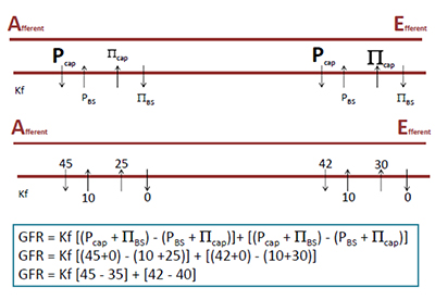
Figure 7. Top: Shows the different forces acting to favour and oppose filtration along the glomerular capillary Bottom: Shows the magnitude of the forces affecting filtration (all in millimetres of mercury, mmHg)
Although filtration is often considered to be a passive process it does indirectly require energy. The energy is needed to constrict the efferent arterioles and for the heart to eject blood from the ventricle and create the pressure to drive filtration.
What volume does the glomerulus filter?
The volume of filtrate produced by the glomeruli is huge; a result of the large surface area, a highly permeable filter, forces favouring filtration and the high cortical blood flow. In health 125ml/min is produced (the GFR), equivalent to 180L/day. Given that a normal adult’s plasma volume is approximately 3 litres, this is the equivalent of the entire plasma volume being filtered by the kidneys 60 times daily. Clearly, the vast majority of this must be reabsorbed further along the nephron otherwise life- threatening hypovolaemia would occur within 15 minutes! In fact, over 99% of the glomerular filtrate is subsequently reabsorbed as it passes along the nephron.
What is the filtration fraction?
This is the fraction of plasma entering the glomerular capillary that is filtered. As previously explained, it is only the plasma component of blood that can take part in filtration. Renal plasma flow can be calculated if renal blood flow and the haematocrit are known. Once renal plasma flow is known the percentage of this that is filtered i.e. the filtration fraction can be calculated.
Renal blood flow x (1 – haematocrit) = Renal plasma flow
e.g. 1000ml/min x (1 – 0.4) = 1000ml/min x 0.6 = 600ml/min
Filtration fraction = GFR / renal plasma flow = 125/600 = 20%
REGULATION OF GLOMERULAR BLOOD FLOW
Autoregulation
Systemic blood pressure and the relative distribution of cardiac output are continuously varying under the control of the autonomic nervous system. If these were allowed to cause alterations in renal blood flow, then the GFR would be unpredictable. In order to prevent this, renal blood flow is kept constant across a wide range of perfusing pressures i.e. renal blood flow is autoregulated, and the GFR kept almost constant (see figure 8). The mechanisms which contribute to this autoregulation are thought to be:
- Myogenic
- Alterations in afferent arteriole blood flow rate alter the tension (stretch) of the arteriole wall, and if left unchecked would alter glomerular blood flow
- An increase in afferent arteriole blood flow would tend to increase glomerular blood flow and increases arteriole wall tension
- This leads to reflex constriction of the afferent arteriole, increasing resistance to blood flow
- As a result the glomerular blood flow returns to normal
- The opposite changes occur when the afferent arteriole tension falls, i.e. a reflex relaxation of the arteriole lowering resistance to blood flow resulting in an increase in glomerular perfusion
- Tubuloglomerular feedback
- Alterations in GFR due to changes in glomerular perfusion pressure will lead to an alteration in the composition of the fluid delivered to the macula densa region of the nephron tubule
- The macula densa senses these changes and acts to alter afferent arteriole tone to vary glomerular perfusion pressure to return the GFR to normal
- The precise details of what is sensed in the tubular fluid are still not known
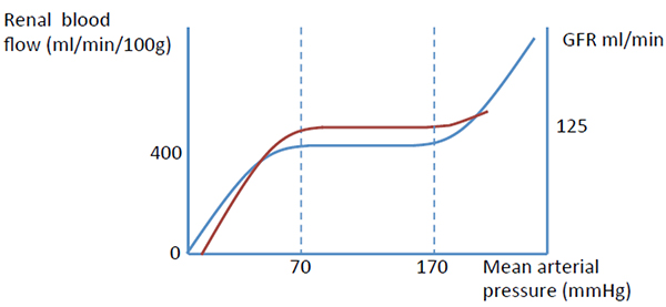
Figure 8. Graph showing the almost constant renal blood flow and GFR across a range of perfusion pressures
Autoregulation aims to ensure that changes in blood pressure do not alter renal blood flow or GFR. There are also a number of other factors that affect renal blood flow and GFR.
The sympathetic nervous system (SNS)
SNS activation causes widespread vasoconstriction mediated by noradrenaline acting on α1 adrenoreceptors on blood vessel smooth muscle cells. The afferent and efferent arterioles receive sympathetic innervation, and both constrict in response to increased SNS activity. This results in a significantly reduced renal blood flow. However, glomerular perfusion pressure is maintained due to greater constriction of the efferent arterioles. Overall the GFR only drops a little (see Figure 9b)
The renin-angiotensin- aldosterone system
The afferent arteriole wall contains some specialised ‘granular’ cells which secrete the proteolytic hormone renin. Renin release is stimulated by:
- Decreased afferent arteriole wall tension
- SNS activity acting on β1 adrenoreceptors of the granular cells
- Decreased sodium and chloride delivery to the macula densa (similar to tubuloglomerular feedback)
Renin converts angiotensinogen to angiotensin 1 which is subsequently converted to angiotensin 2 by angiotensin converting enzyme (ACE). Angiotensin 2 causes greater constriction of efferent than afferent arterioles. Overall the GFR is maintained through an increase in filtration fraction.
Renal prostaglandins
Prostaglandins are produced from arachidonic acid within the kidney when renal blood flow is compromised, for example during increased SNS activity. Prostacyclin (PGI2) acts to cause afferent arteriole vasodilatation to maintain glomerular blood flow and GFR.
Atrial natriuretic peptide (ANP)
ANP is stored in cardiac atrial cells and released in response to increased atrial stretch i.e. an expansion in circulating volume. One of the actions of ANP is to cause vasodilatation of the afferent arterioles and increase GFR.
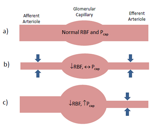
Figure 9 a) Normal afferent and efferent arteriole tone
b) Afferent and efferent vasoconstriction (e.g. SNS activity or Angiotensin 2) resulting in reduced RBF, but maintained Pcap and GFR
c) Efferent vasoconstriction with a normal afferent tone (e.g. SNS activity with action of renal prostacyclin) resulting in reduced RBF but a well maintained Pcap and GFR
SUMMARY
- The kidneys are high specialised organs with an intricate and highly ordered structure
- The basic functional unit of the kidney is the nephron
- The kidneys play a vital role in fluid and electrolyte homeostasis within the body. The first step in this process is ultrafiltration of plasma at the glomerulus which requires careful control of glomerular blood flow
- The vast majority of the fluid that is filtered at the glomerulus is reabsorbed further along the nephron
Part 2 of this tutorial will focus on how re-absorption of this fluid occurs and is controlled in order to maintain a constant extracellular fluid volume and osmolality.
ANSWERS
- F, F, T, F
The kidneys are situated at the level of T12 – L3 but are retroperitoneal organs measuring approximately 12cm from end to end. Each kidney has two clearly distinguishable regions; the cortex and the medulla. The abdominal aorta is slightly to the left of the midline while the IVC is slightly to the right; therefore the left kidney has a slightly shorter renal artery but longer renal vein. - T, F, F, T
There are two different types of nephrons; cortical and juxtamedullary. Regardless of the type of nephrons, all of the glomeruli are within the cortex of the kidney. The juxtaglomerular apparatus is situated after the loop of Henle, i.e. in the distal tubule. All of the collecting ducts pass through the medulla and drain the urine that is produced into the calyces. - T, F, F, F
Renal blood flow is around 400ml/100g/min, which higher than either coronary or cerebral blood flow. This blood flow is unevenly distributed with the vast majority supplying the cortex. The high flow rate and preferential distribution to the cortex is necessary to drive glomerular filtration, rather than to meet metabolic demands. - T, T, T, F
In health the glomerular filtration rate is 125ml/min. The main barrier to glomerular filtration is the glomerular basement membrane. This is made from connective tissue which is negatively charged and so tends to oppose the filtration of negatively charged molecules e.g. plasma proteins. Small molecules such as electrolytes, urea and glucose are freely filtered so that the ultrafiltrate produced by the glomerulus has the same osmolality as plasma.
This work by WFSA is licensed under a Creative Commons Attribution-NonCommercial-NoDerivitives 4.0 International License. To view this license, visit https://creativecommons.org/licenses/by-nc-nd/4.0/