Regional Anaesthesia

KEY POINTS
- Ultrasound-guided interscalene brachial plexus block can provide dense surgical anaesthesia, and/or intra- and postoperative analgesia, for shoulder surgery.
- Patient selection is particularly important for interscalene block safety, since the frequently associated phrenic nerve block can cause significant respiratory distress in patients with preexisting respiratory disease.
- Practitioners should use dynamic ultrasound scanning and colour Doppler to correctly identify the brachial plexus roots in the interscalene groove and to avoid inadvertent intravascular injection.
- Tracking the needle tip, assessing the local anaesthetic spread, and maintaining a cautious approach to the nerve roots are fundamental to reducing the risk of complications such as nerve injury or local anaesthetic toxicity.
INTRODUCTION
The ultrasound-guided interscalene block (ISB) has a faster onset time and longer duration than the nerve-stimulation technique, and it produces surgical anaesthesia more reliably for the same volume of local anaesthetic.1 Likewise, lower volumes of local anaesthetic are needed for an effective block.2 An ultrasound-guided technique can also decrease the incidence of hemidiaphragmetic paresis.3 However, the incidence and severity of postoperative neurological symptoms are similar between both techniques.4 A periplexus approach to the ultrasound-guided ISB, with deposition of local anaesthetic in the potential space between middle scalene (MS) muscle and brachial plexus sheath, produces the same quality of block as the classic intraplexus approach (where the needle tip is placed between individual nerve roots).5,6 Performing the block outside the brachial plexus sheath under ultrasound visualization, using a periplexus approach, may reduce the risk of nerve injury.7
ANATOMY
Brachial Plexus
- The brachial plexus innervates the shoulder and upper limb (Figure 1). The roots originate from the ventral rami of cervical (C) spinal nerves C5 through C8 and the first thoracic spinal nerve, T1 (Figure 2).8 Variations include having a ‘‘prefixed’’ plexus that originates from C4 through C8 or a ‘‘postfixed’’ plexus that originates from C6 through C8 and T1 to T2.9 A study of anatomic variation in human fetuses identified a ‘‘prefixed’’ plexus in 25.5% of cases and a ‘‘postfixed’’ plexus in 2.5% of cases.10
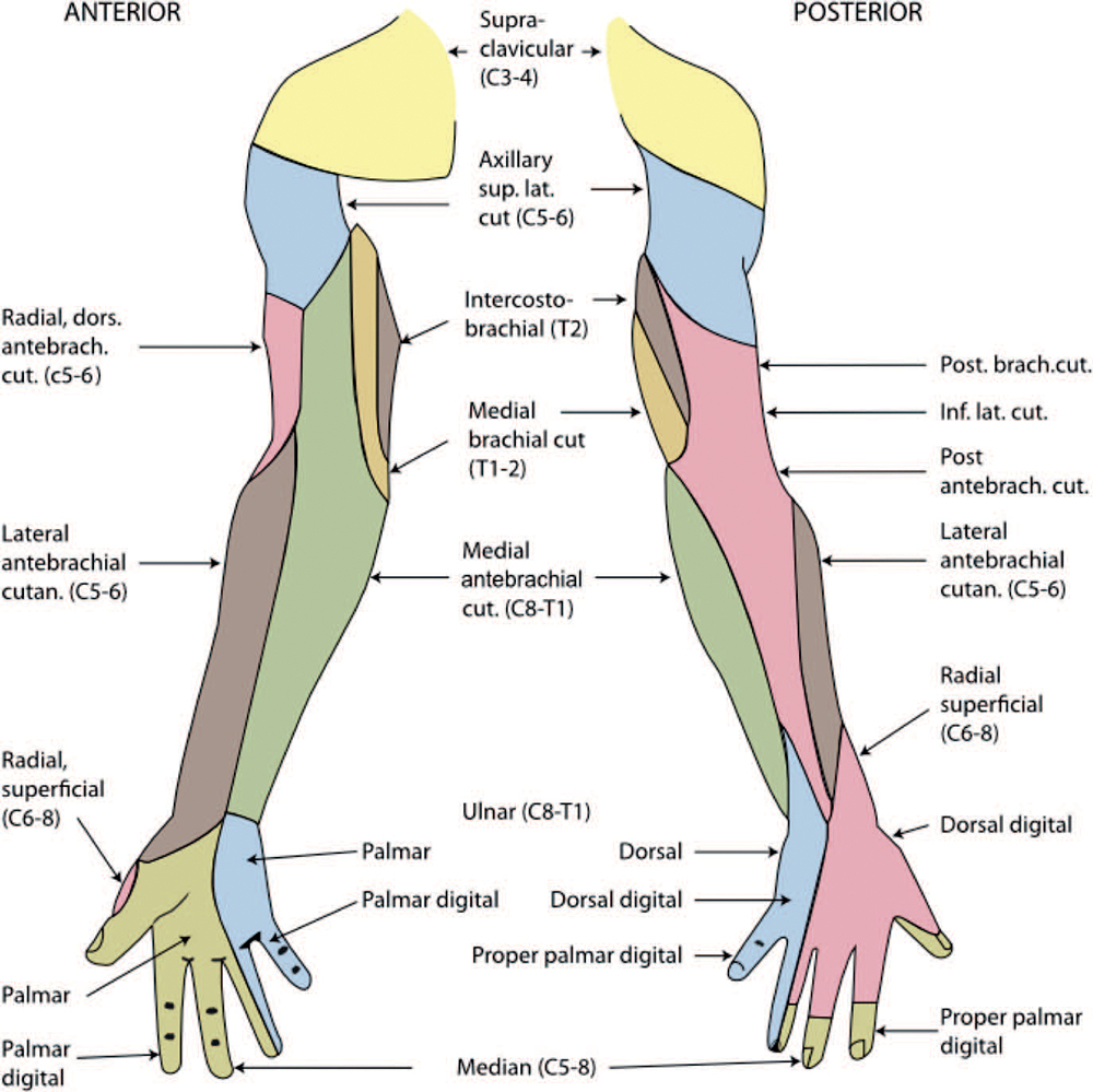
Figure 1. Sensory innervation of the upper extremity. Source: Henry Vandyke Carter [Public domain], via Wikimedia Commons: https://upload. wikimedia.org/wikipedia/commons/d/de/Gray812and814.PNG.
- The brachial plexus’s upper roots are typically found in the ‘‘interscalene groove’’ between the anterior scalene (AS) and MS muscles, although the C5 root may be anterior to the AS and both C5 and C6 roots may pass through the AS.11
- The C6 nerve root often bifurcates into 2 fascicles, within a common epineurium, before forming the upper trunk with the C5 nerve root.12
Adjacent Structures
- The cervical plexus provides sensory innervation to the ‘‘shoulder cape’’ via the supraclavicular nerve (C3-4).
- The cervical plexus provides motor innervation to the ipsilateral diaphragm via the phrenic nerve (C3-5). The phrenic nerve passes between the AS and MS, and then descends on the anterior surface of the AS into the thoracic cavity.
- The recurrent laryngeal nerve provides motor innervation to all but one of the intrinsic laryngeal muscles, the cricothyroid muscle, which is innervated by a branch of the superior laryngeal nerve.
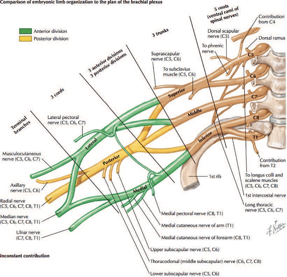
Figure 2. Anatomic representation of the embryonic limb organization of the brachial plexus. Copyright Elsevier Netter Images. Used with permission.
- The sympathetic nerve fibres innervating the head exit the spinal cord at upper thoracic levels (T1) before entering the sympathetic chain and ascending to the superior cervical ganglion.
- The long thoracic and dorsal scapular nerves originate from the proximal brachial plexus and frequently pass through the MS at the same level and slightly posterior to the C6 nerve root.13
- The carotid and vertebral arteries and the internal and external jugular veins follow a course perpendicular, and in proximity, to the brachial plexus nerve roots.
- The subclavian artery runs parallel to the brachial plexus nerve roots at the level of C8 and T1. At the level of the trunks, the brachial plexus is posterior and cephalic in relation to the artery.14
- The transverse cervical artery, which arises from the thyrocervical trunk of the subclavian artery, crosses anterior to the scalene muscles.
- The apex of the lungs extends into the root of the neck, reaching up to 4 cm above the middle third of the clavicle.
Ultrasound
- At the interscalene groove, the C5-7 nerve roots are better visualized than C8 and T1 due to the depth and cephalo-caudad position of each nerve root. The brachial plexus’s roots appear as round homogenous hypoechoic (dark) structures (Figure 3), not the classic honeycomb pattern that is seen distally in the brachial plexus. This difference is due to the high density of conducting axons and relatively little hyperechoic connective tissue found elsewhere in the body. Differentiating the nerve roots from other adjacent hypoechoic structures, such as blood vessels, requires careful attention and can be aided by the routine use of colour Doppler. It is also more difficult to capture all the brachial plexus nerve roots in the same image due to their varied angulation upon exiting the spinal cord.
- The brachial plexus can be readily identified at the supraclavicular level where the trunks run parallel to each other and are most frequently lateral to the subclavian artery, though anatomical variations exist where the superior trunks may be anterior or medial the artery (Figure 4).14 The roots can then be located using the ‘‘traceback’’ method, sliding the transducer cephalad while maintaining visualization of the plexus up to the interscalene groove (Figure 5).
- The ‘‘stoplight sign’’ is a frequently cited name for the typical sonographic appearance of 3 root structures aligned vertically in the interscalene groove. The 3 structures most often represent, from cephalad to caudal: the C5 root, the upper fascicle of C6, and the lower fascicle of C6.12
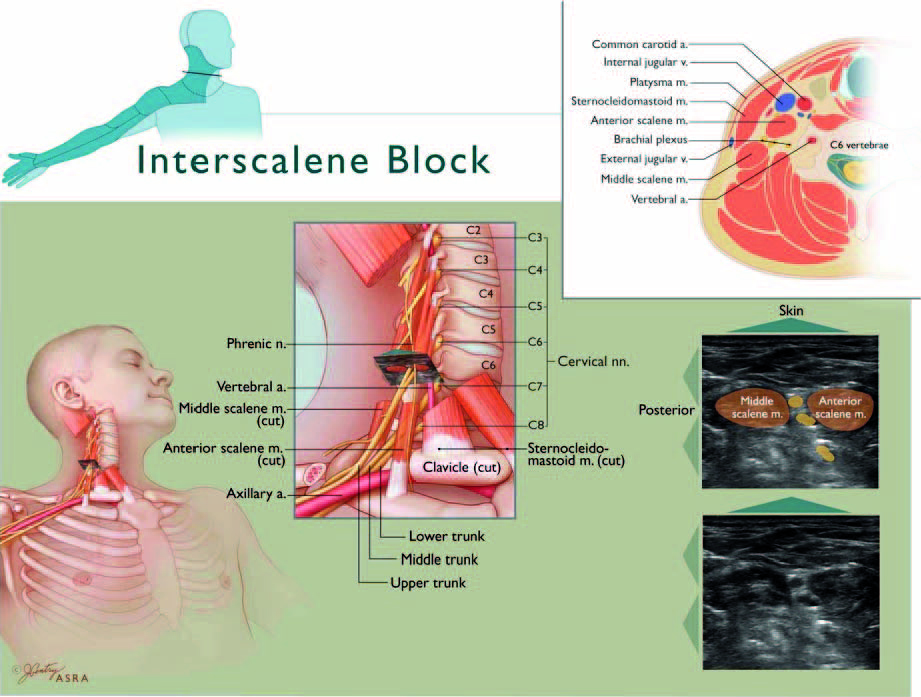
Figure 3. Anatomic representation and ultrasound image of the interscalene groove. Copyright American Society of Regional Anesthesia and Pain Medicine. Used with permission.
- Identifying the cervical level with ultrasound is based on the difference between C6 (Figure 6) and C7 (Figure 7) transverse process anatomy. The C7 transverse process is identified by the small anterior tubercle on ultrasound, and it can be compared to C6 by sliding the transducer between adjacent vertebrae (Figure 8).15
- The long thoracic and dorsal scapular nerves appear as discrete hyperechoic structures with a hypoechoic centre within the MS muscle.13
INTERSCALENE BLOCK
Indications
The ISB provides reliable anaesthesia and analgesia to the shoulder and proximal arm because it consistently blocks the C5-6 nerve roots.16,17 With higher volumes of local anaesthetic, the block also affects the superficial cervical plexus including the supraclavicular nerve, providing a sensory block to the ‘‘shoulder cape.’’
Contraindications
General contraindications to regional anaesthesia and specific contraindications to the ISB are presented in Table 1. Determining whether or not the ISB is contraindicated in a patient requires a thorough understanding of the side effects and complications that are specific to this block.
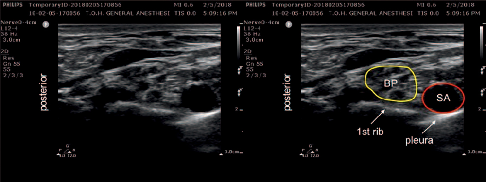
Figure 4. Ultrasound image of supraclavicular view of the brachial plexus (BP) and subclavian artery (SA).
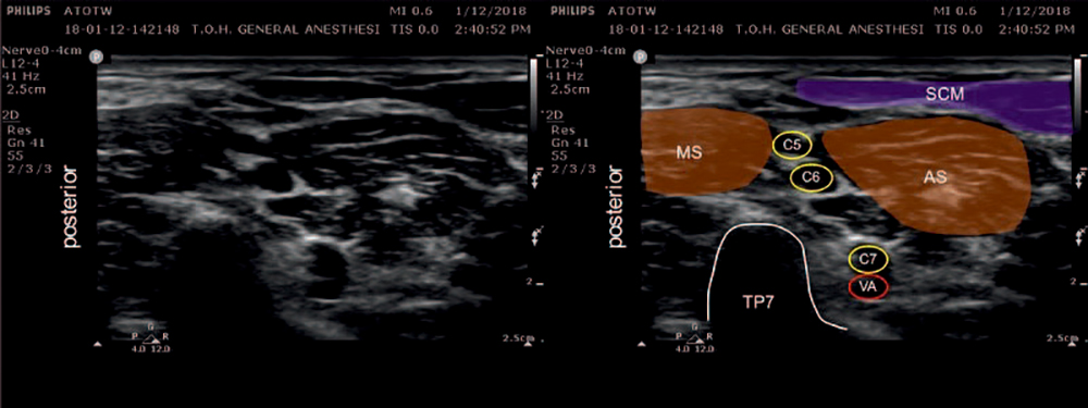
Figure 5. Ultrasound image of the interscalene groove. AS indicates anterior scalene; MS, middle scalene; SCM, sternocleidomastoid; TP, transverse process of the 7th cervical vertebra; C5-7, 5th to 7th cervical nerve roots; And VA, vertebral artery.
Side Effects and Complications
The side effects and complications related to ISB are presented in Table 2. Compared to other peripheral nerve blocks, the ISB carries the highest risk of neurological injury. The estimated rate of occurrence of neuropathy after ISB is 2.84 per 100 patients.18 Depending on the volume used, the incidence of ipsilateral phrenic nerve block is up to 100%.19 Contralateral or severe bilateral respiratory impairments are thus contraindications to ISB (eg, chest trauma, pneumonectomy, pneumothorax, severe chronic obstructive pulmonary disease). Similarly, ISB is contraindicated in the presence of contralateral phrenic nerve deficit, since blocking the functional phrenic nerve may result in severe respiratory distress or respiratory arrest. Preexisting unilateral vocal cord paralysis also limits the use of ISB on the contralateral side because of the risk of recurrent laryngeal nerve block which may precipitate glottic obstruction.
TECHNIQUE
Equipment
- Standard physiologic monitors (electrocardiogram, pulse oximeter, noninvasive blood pressure)
- Resuscitation equipment including 20% lipid emulsion (Intralipid)
- Skin antiseptic, preferably chlorhexidine/alcohol based
- Sterile gloves, drapes, and transducer cover
- Ultrasound machine with a high-frequency linear transducer
- Short-bevel regional block needle (eg, 50 mm, 22 gauge)
- Normal saline or 5% dextrose solution (if using nerve stimulation in addition to ultrasound guidance)
- Short-acting local anaesthetic such as lignocaine for skin infiltration (1% or 2%)
- Local anaesthetic solution for plexus block
Safe and successful regional anaesthesia also requires a skilled assistant.
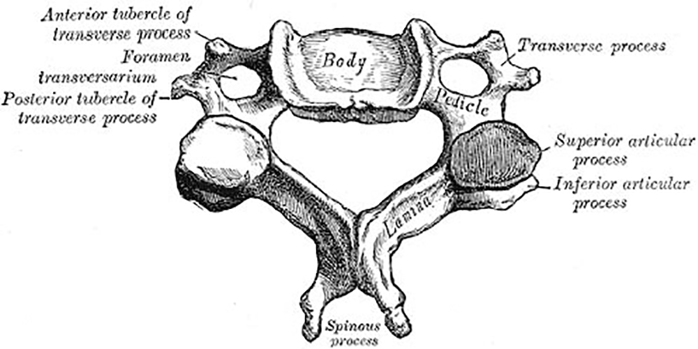
Figure 6. Cervical vertebrae anatomy. Source: Henry Vandyke Carter [Public domain], via Wikimedia Commons: https://upload.wikimedia.org/wikipedia/commons/3/3c/Gray84.png
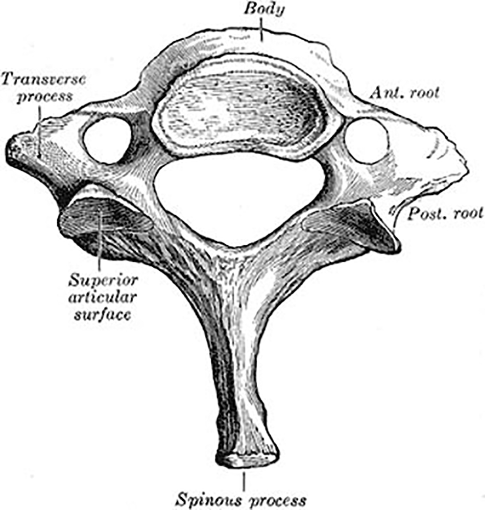
Figure 7. Seventh cervical vertebra anatomy. Source: Henry Vandyke Carter [Public domain], via Wikimedia Commons: https://upload.wikimedia.org/wikipedia/commons/c/cf/Gray89.png.
Positioning
The patient can be positioned supine with their head rotated towards the contralateral side, or in the lateral decubitus position with the block side up. The operator can position themselves at the patient’s side or at the head of the bed. Proper bed height and ergonomics are key to the safe conduct of regional anaesthesia.
Approach and Scanning
- Gain intravenous access, apply standard monitoring, and perform institutional preprocedural safety checklist (see Table 3).20
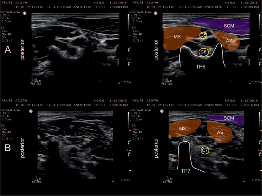
Figure 8. A. Ultrasound image of C6 transverse process. B. Ultrasound image of C7 transverse process. AS indicates anterior scalene; MS, middle scalene; SCM, sternocleidomastoid; C5-7, 5th to 7th cervical nerve roots; and TP6-7; 6th and 7th cervical vertebrae transverse processes.
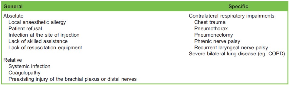
Table 1. Contraindications to the Interscalene Block
- Position the patient and perform skin preparation of the neck and supraclavicular area with chlorhexidine/alcohol before applying sterile drapes and covering the transducer with a sterile cover.
- Position the transducer in the supraclavicular fossa just cephalad and parallel to the clavicle to identify the subclavian artery, first rib, lung pleura and the brachial plexus trunks.
- Using the ‘‘traceback’’ method, follow the nerve trunks back up to the interscalene groove by sliding the transducer cephalad while maintaining visualization of the plexus.
- Once the brachial plexus nerve roots have been identified in the interscalene groove, confirm the cervical level based on transverse process anatomy. Use colour Doppler to identify vascular structures, paying specific attention to avoid the vertebral artery and the transverse cervical artery.
- When visualizing the ‘‘stoplight sign,’’ the C6 nerve root’s fascicles are correctly identified by tracing the nerve root proximal to its bifurcation where the nerve exits the intervertebral foramen.
- Confirm appropriate probe position before attempting needle puncture. The probe should be oriented transversely across the lateral neck, overlying the sternocleidomastoid and scalene muscles, at approximately the level of the cricoid cartilage.
Block Conduct—Periplexus Interscalene Block
- Confirm the identity of critical landmarks: AS, MS, and the C5 and C6 roots within the interscalene groove.
- Infiltrate the skin with 1 to 3 mL of local anaesthetic.
- Advance the block needle in plane through skin and MS towards the C5 and C6 roots in the interscalene groove (Figure 9).
- As the needle traverses MS, be careful to avoid injuring the long thoracic and dorsal scapular nerves.
- Advance the needle through the MS and stop at the border of its fascial layer with the hyperechoic fascia covering the brachial plexus.
- A subtle ‘‘pop’’ sensation may be appreciated when the needle tip enters the interscalene groove. The needle tip does not need to pierce the brachial plexus sheath (periplexus approach). This approach is advised to reduce the risk of nerve injury.
- Aspirate to rule out an intravascular needle position and inject 1 mL of saline or 5% dextrose to confirm deposition between the fascial layers.
- To avoid intraneural injection, only inject with low pressures, observe the nerve roots to rule out nerve expansion (which indicates intraneural injection), and communicate with the patient to rule out paresthesias during needle advancement.
- If the injectate spreads posteriorly along the needle tract within MS, advance another 1 to 2 mm, being careful to avoid piercing the nerve roots with the needle.
- Correct injectate deposition will appear on ultrasound as the plexus being pushed medially with hypoechoic spread adjacent to the nerve roots (Figure 9).
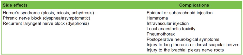
Table 2. Side Effects and Complications of the Interscalene Block
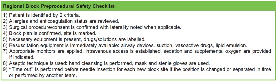
Table 3. Example of a Preblock Safety Checklist, from Mulroy et al20
- For block success, local anaesthetic must be visualized within the correct tissue plane of the interscalene groove. However, complete circumferential spread around the plexus is not necessary. Our practice is to scan proximally and distally from the injection site several times over the course of the injection to confirm correct deposition not only in the plane of our needle, but also along the course of the plexus.
- Once satisfied with needle position, switch to local anaesthetic. Inject the total desired volume in 3- to 5-mL increments, while continuing to aspirate intermittently to rule out an intravascular injection and confirming low injection pressures.
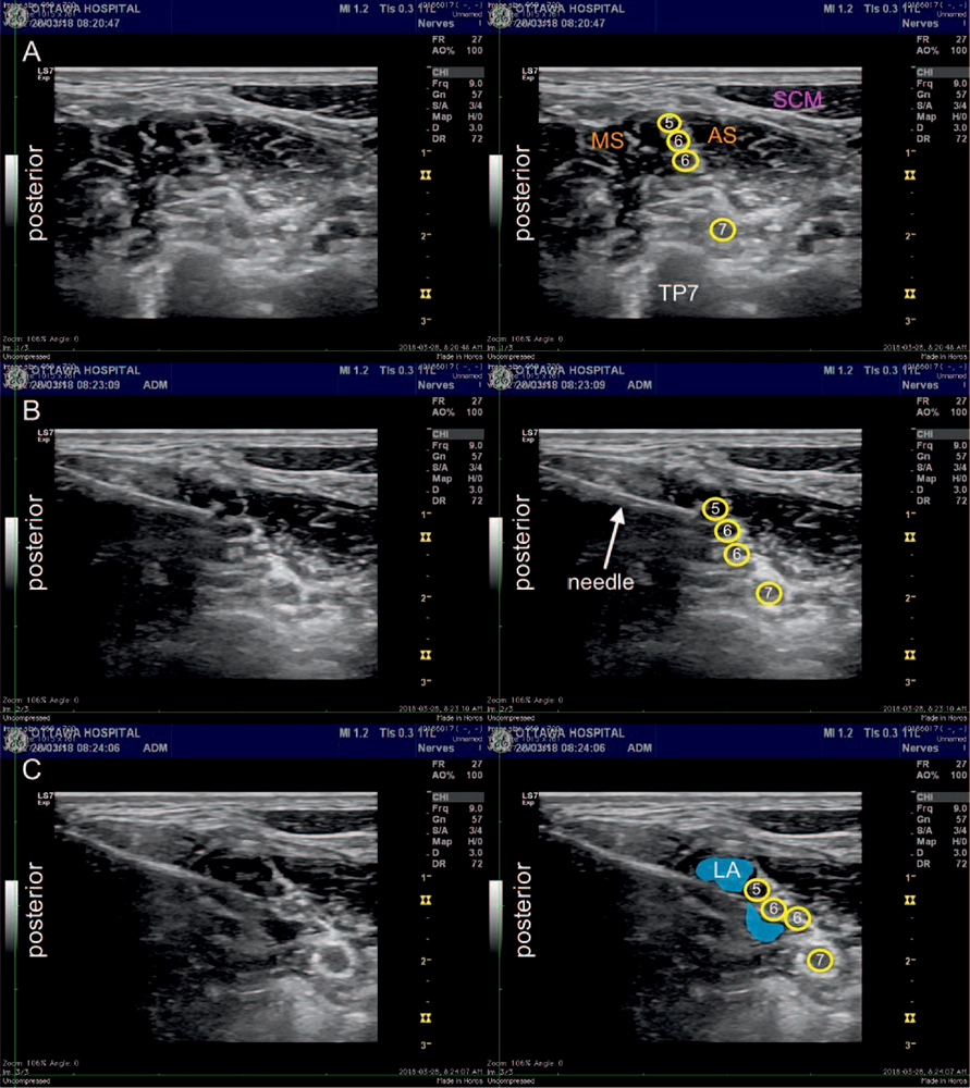
Figure 9. A. Interscalene block preinjection. B. Interscalene block with needle tip outside the brachial plexus sheath. C. Interscalene block final local anesthetic spread (blue) around brachial plexus. AS indicates anterior scalene; MS, middle scalene; SCM, sternocleidomastoid; TP7, transverse process of 7th cervical vertebra; yellow circles 5 to 7, brachial plexus nerve roots; LA, local anaesthetic; and blue, local anaesthetic spread. Note: The images demonstrate the common finding of a C6 nerve root that has divided into upper and lower fascicles.
- Usual doses of the block are 15 to 30 mL of 0.5% bupivacaine or ropivacaine consistent with the overall local anaesthetic dosing of less than 2.5 mg/kg.
Continuous Catheter
A perineural catheter can be placed to extend the duration of postoperative analgesia. Compared to single shot ISB alone, the continuous infusion reduces pain on the first postoperative day.21 Some institutions, including our own, have set up ambulatory peripheral nerve block catheter programs. Most of our patients undergoing rotator cuff repair and shoulder arthroplasty are managed with ambulatory interscalene catheters. Numerous peripheral nerve block catheter kits are commercially available, as well as infusion pumps designed for outpatients. Our standard protocol is to infuse the catheter with 0.2% ropivacaine at 5 mL per hour, with a patient-controlled bolus option of 5 mL every 30 minutes. Patients are discharged home with 250-mL bags of ropivacaine, which lasts up to 48 hours depending on bolus usage. The technical details of interscalene catheter insertion are beyond the scope of this tutorial. The success of an ambulatory catheter program depends greatly on appropriate patient selection, patient training on anticipated effects and potential complications, and a robust follow-up system for troubleshooting. For further reading on peripheral nerve blocks for ambulatory surgery, we suggest the reviews by Salinas and Joseph22 and by Swenson et al.23
SUMMARY
- The ISB consistently blocks the C5-6 nerve roots, providing reliable anaesthesia and analgesia to the shoulder and proximal arm.
- Common side effects of ISB include hemidiaphragm paresis and recurrent laryngeal nerve block. These side effects are well tolerated in most patients but can cause severe respiratory distress in the presence of respiratory disease or contralateral lesions. Therefore, patient screening and selection is of paramount importance for ISB safety.
- Ultrasound identification of the nerve roots at the interscalene groove is facilitated by tracing the brachial plexus proximally from the supraclavicular level.
- Tracing individual nerve roots proximally to the vertebrae allows for cervical-level identification by comparing transverse process anatomy: the C7 transverse process does not have a prominent anterior tubercle.
- We advocate the use of a periplexus approach: position the needle tip in the potential space between the MS and brachial plexus sheath for safe and effective local anaesthetic deposition.
ACKNOWLEDGEMENTS
Dr Christopher Ramnanan kindly reviewed the anatomy section.
REFERENCES
- Kapral S, Greher M, Huber G, et al. Ultrasonographic guidance improves the success rate of interscalene brachial plexus blockade. Reg Anesth Pain Med. 2008;33(3):253-258V
- McNaught A, Shastri U, Carmichael N, et al. Ultrasound reduces the minimum effective local anaesthetic volume compared with peripheral nerve stimulation for interscalene block. Br J Anaesth. 2011;106(1):124-130.
- Ghodki PS, Singh ND. Incidence of hemidiaphragmatic paresis after peripheral nerve stimulator versus ultrasound guided interscalene brachial plexus block. J Anaesthesiol Clin Pharmacol. 2016;32(2):177-181.
- Liu SS, Zayas VM, Gordon MA, et al. A prospective, randomized, controlled trial comparing ultrasound versus nerve stimulator guidance for interscalene block for ambulatory shoulder surgery for postoperative neurological symptoms. Anesth Analg. 2009;109(1):265-271.
- Spence BC, Beach ML, Gallagher JD, Sites BD. Ultrasound-guided interscalene blocks: understanding where to inject the local anaesthetic. Anaesthesia 2011;66(6):509-514.
- Maga J, Missair A, Visan A, et al. Comparison of outside versus inside brachial plexus sheath injection for ultrasoundguided interscalene nerve blocks. J Ultrasound Med. 2016;35(2):279-285.
- Szerb JJ, Greenberg JL, Kwofie MK, et al. Histological confirmation of needle tip position during ultrasound-guided interscalene block: a randomized comparison between the intraplexus and the periplexus approach. Can J Anesth. 2015;62(12):1295-1302.
- Hansen JT. Netter’s Clinical Anatomy. 3rd ed. Philadelphia, PA: Saunders/Elsevier; 2014.
- Ellis H, Lawson A. Anatomy for Anaesthetists. 9th ed. Chichester, West Sussex, UK: John Wiley & Sons Ltd; 2014.
- Uysal II, Sxeker M, Karabulut AK, et al. Brachial plexus variations in human fetuses. Neurosurgery. 2003;53(3):676-684.
- Harry WG, Bennett JDC, Guha SC. Scalene muscles and the brachial plexus: anatomical variations and their clinical significance. Clin Anat. 1997;10(4):250-252.
- Franco CD, Williams JM. Ultrasound-guided interscalene block reevaluation of the stoplight sign and clinical implications. Reg Anesth Pain Med. 2016;41(4):452-459.
- Hanson NA, Auyong DB. Systematic ultrasound identification of the dorsal scapular and long thoracic nerves during interscalene block. Reg Anesth Pain Med. 2013;38(1):54-57.
- Van Geffen GJ, Moayeri N, Bruhn J, Scheffer GJ, Chan VW, Groen GJ. Correlation between ultrasound imaging, crosssectional anatomy, and histology of the brachial plexus: a review. Reg Anesth Pain Med. 2009;34(5):490-497.
- Martinoli C, Bianchi S, Santacroce E, Pugliese F, Graif M, Derchi LE. Brachial plexus sonography: a technique for assessing the root level. Am J Roentgenol. 2002;179(3):699-702.
- Vester-Andersex T, Christiansen C, Hansen A, Sørensen M, Meisler C. Interscalene brachial plexus block: area of analgesia, complications and blood concentrations of local anesthetics. Acta Anaesthesiol Scand. 1981;25(2):81-84.
- Lanz E, Theiss D, Jankovic D. The extent of blockade following various techniques of brachial plexus block. Anesth Analg. 1983;62(1):55-58.
- Brull R, McCartney CJL, Chan VWS, El-Beheiry H. Neurological complications after regional anesthesia: contemporary estimates of risk. Anesth Analg. 2007;104(4):965-974.
- Riazi S, Carmichael N, Awad I, Holtby RM, McCartney CJL. Effect of local anaesthetic volume (20 vs 5 ml) on the efficacy and respiratory consequences of ultrasound-guided interscalene brachial plexus block. Br J Anaesth. 2008;101(4):549 556.
- Mulroy MF, Weller RS, Liguori GA. A checklist for performing regional nerve blocks. Reg Anesth Pain Med. 2014;39(3):195-199.
- Mariano ER, Afra R, Loland VJ, et al. Continuous interscalene brachial plexus block via an ultrasound-guided posterior approach: a randomized, triple-masked, placebo-controlled study. Anesth Analg. 2009;108(5):1688-1694.
- Salinas FV, Joseph RS. Peripheral nerve blocks for ambulatory surgery. Anesthesiol Clin. 2014;32(2):341-355.
- Swenson JD, Cheng GS, Axelrod DA, Davis JJ. Ambulatory anesthesia and regional catheters: when and how. em>Anesthesiol Clin. 2010;28(2):267-280.
This work by WFSA is licensed under a Creative Commons Attribution-NonCommercial-NoDerivitives 4.0 International License. To view this license, visit https://creativecommons.org/licenses/by-nc-nd/4.0/