Intensive Care Medicine

This tutorial will present several vignettes of clinical scenarios. Attempt to answer the questions posed with each one before reading the discussion section.
Case 1
An otherwise fit and well 60-year-old man presents for elective varicose vein surgery. The junior doctor on the ward orders a chest X-ray as the patient has recently had a chest infection – he is now asymptomatic.
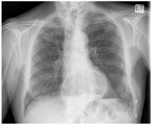
Figure 1. Chest X-ray
What coincidental abnormality does this chest X-ray show? Would this change your preoperative management? How would you alter the anaesthetic you give him?
Case 2
A previously fit 30-year-old man is brought to the Emergency Department with a Glasgow Coma Score of 3/15 having been knocked off his motorcycle. He is being ventilated using a bag-valve-mask, has a heart rate of 80min-1 and a blood pressure of 150/80mmHg. You immobilise his cervical spine, intubate him and take him for an urgent CT head scan (Figure 2). No other significant injuries have been found clinically or radiologically.
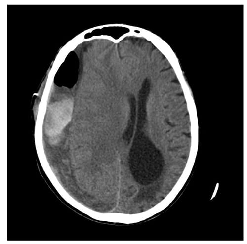
Figure 2. CT head scan
What does his CT head show? How would you manage this patient?
Case 3
You are on a ward assessing patients for surgery when a patient in a nearby cubicle collapses. The emergency team is called but you are first on the scene. The patient is 65-years-old and is awaiting elective surgery. He responds to painful stimulus, a weak radial pulse is palpable and the nurse has recorded a blood pressure of 70/45mmHg. The ECG is shown below. 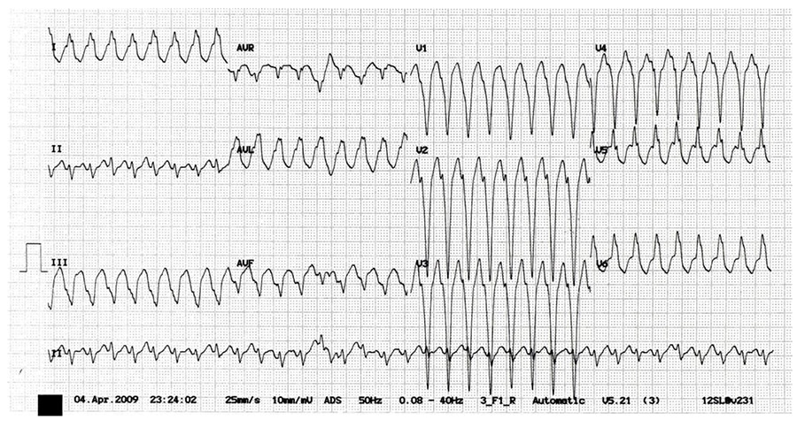
What abnormality is shown on the ECG? How would you initially manage this patient? How would you manage this patient if they were not compromised by the arrhythmia? How would you decide if this was a ventricular or supra-ventricular rhythm? Would it change your initial management?
DISCUSSION
Case 1
This chest Xray shows a large hiatus hernia. This can be seen behind the heart as a globular structure with a fluid level within it. The Xray film must be adequately penetrated to show structures behind the heart – i.e. you should just be able to see the thoracic vertebral bodies behind the heart. Hiatus hernias arise due to a defect in the diaphragm allowing part or all of the stomach to pass into the thorax. The patient may have symptoms of dyspepsia, belching, chest pain (which can mimic cardiac ischaemia) or frank reflux. Hiatus herniae are relevant in anaesthesia due to the increased risk of reflux and subsequent aspiration. Pulmonary aspiration of gastric contents carries a significant morbidity and mortality. Injury from aspiration of gastric contents results from chemical pneumonitis, mechanical obstruction from particulate material and bacterial contamination. The morbidity is related to the pH and the particle size of the aspirate. Patients can be at greater risk of reflux due to a variety of factors (Table 1).
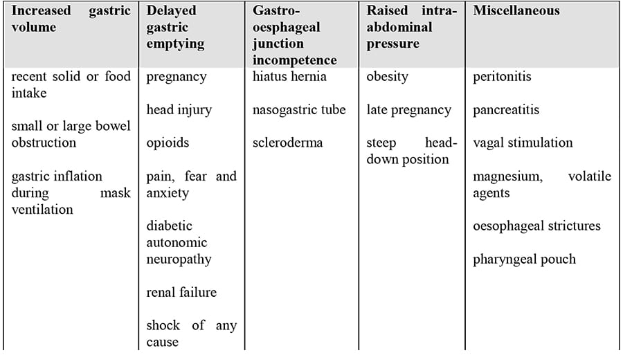
Table 1. Risk factors for gastro-oesophageal reflux
In order to minimise the risk of reflux, aspiration and resultant morbidity, anaesthetic practitioners should consider the following: Preoperative care
- Patients should be appropriately fasted (2 hours for clear fluids, 6 hours for food/milk).
- Patients should be questioned about the presence of symptoms such as heartburn or reflux or previous diagnosis of hiatus hernia.
- Consideration should be given to the use of premedicating agents to reduce the volume and increase the pH of gastric contents (see Table 2).
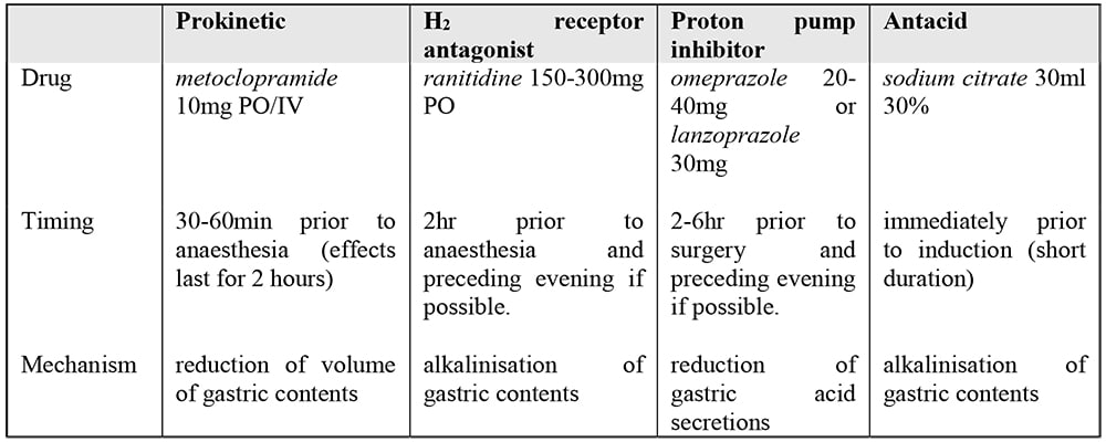
Table 2. Summary of premedicant drugs available for patients with reflux oesophageal disease
In the presence of significant symptoms of reflux, the general anaesthesia technique should include intubation, inserted as part of a rapid sequence induction. Significant reflux symptoms should be considered as:
- frequent (daily to weekly) reflux – felt as acid in the mouth or burning in the oesophageal area.
- reflux related to position (lying flat or bending over).
For some patients, who give a history of infrequent reflux, you may feel that intubation is not necessary. For these patients one or more of the premedicant drugs shown in Table 2 may be used to enhance gastric emptying or reduce the acidity of the stomach contents. This decision will also be influenced by the planned procedure and other factors that may make reflux more likely. If you are uncertain whether the reflux history is significant, the safest course of action is to intubate using a rapid sequence induction. Postoperative care
- Muscle relaxant should be adequately reversed and the patient fully awake (able to sustain a head lift for 5 seconds and maintain a firm grip) prior to extubation.
- Extubation should be performed with the patient on their side or sitting up to minimise risk of aspiration at this critical point.
- Make the patient aware of the need to inform future anaesthetic practitioners of their significant aspiration risk.
Case 2
Traumatic brain injuries can be clinically classified on the basis of the GCS: 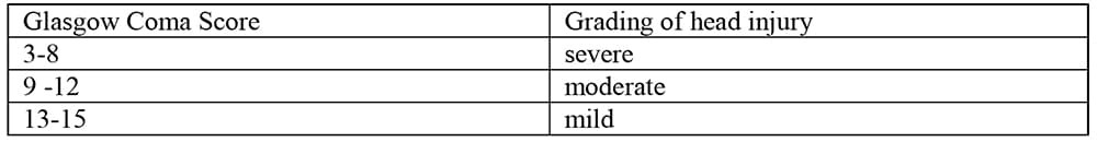 Patients with a GCS less than 9 should be considered to have lost their protective airway reflexes and so should be intubated early. Rapid sequence induction should be used with consideration to use of opioids such as alfentanil to minimise the rise in intracranial pressure associated with intubation. Until proven otherwise, such patients should be assumed to have suffered a cervical spine injury with 3-point immobilisation and in-line stabilisation during intubation. This patient’s CT shows a large right subdural haematoma with significant midline shift indicative of a severe head injury. Subdural haematomas are located between the dura mater lining the brain and the brain itself, with a convex outer edge and a concave inner border. They are usually venous in origin.
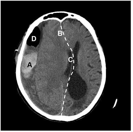
Figure 4. The CT of patient 2 shows a large subdural haematoma (A) with midline shift (B), effacement of the right lateral ventricle (C) and air within the cranial cavity (D) indicating likely cranial fracture
Head injuries can also be classified radiologically by their appearance on a CT scan. 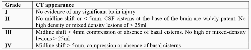 This man should be discussed with a neurosurgeon as definitive management should include drainage of the haematoma. This may involve transfer to a tertiary centre with neurosurgical facilities. Whilst awaiting transfer to theatre or another centre, there are several aspects to his care that can be optimised to limit secondary brain injury. REMEMBER: CPP = MAP – ICP CPP = Cerebral perfusion pressure, MAP = Mean arterial pressure, ICP = intracerebral pressure)
- Normal ICP is 5-12mmHg. In a head injured patient it is reasonable to assume it is 20mmHg and so a mena arterial pressure of 90mmHg is needed to achieve a CPP of 70mmHg.
- Blood pressure should be monitored regularly, and invasively if possible
- If thoracic and lumbar spine have been cleared then patient should be 30° head up to improve cranial venous drainage.
- Endotracheal tubes should be taped in place rather than tied to prevent ties obstructing venous drainage.
- PaCO2 should be maintained in the low normal range (4.5-5kPa).
- Maintain normoglyacemia.
- Maintain oxygen saturations >95%.
- Any seizures should be controlled (e.g. 18mg.kg-1 phenytoin)
A brief period of hyperventilation (to lower the PaCO2) and/or mannitol 0.5g.kg-1 IV can be used to control acute surges in ICP. Hyperventilation should not be sustained as cerebral vasoconstriction may lead to further ischaemia.
FURTHER READING
- Ali B, Drage S. Management of head injuries. Anaesthesia Tutorial of the Week No 46 (March 2007).
- Available at: http://totw.anaesthesiologists.org/2007/03/12/management-of-head-injuries-46/
Case 3
This ECG shows a broad complex tachycardia (the QRS duration is greater than 0.12s or more than 3 small squares) which should be managed according to the European Resuscitation Council’s guidance (Figure 5).
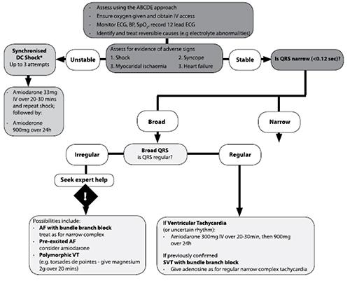
Figure 5. Tachycardia algorithm (By kind permission of the European Resuscitation Council)
As for any acutely unwell patient, management should follow an ABC approach:
- A: Airway support if necessary and administer oxygen (15l.min-1 as available)
- B: Pulse oximetry should be recorded and assessment should be made of his breathing which might be compromised in this situation by pulmonary oedema
- C: Circulation is compromised here – his blood pressure is 70/45. IV access should be obtained.
The cause of this patient’s collapse is his arrhythmia. The management of arrhythmias is determined by two major factors:
- Whether the patient is stable or unstable (as here)
- The nature of the arrhythmia.
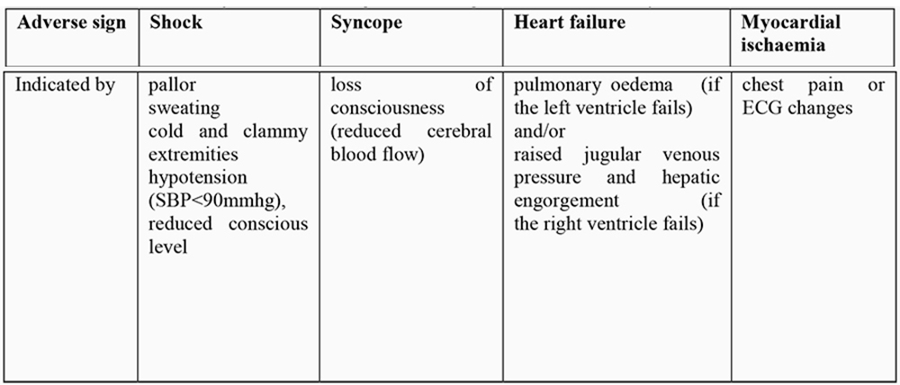
Table 3. Presence of any of the following adverse signs indicates that a patient is unstable
The first step in management is to treat any reversible precipitants such as electrolyte abnormalities (particularly low plasma potassium and low plasma magnesium) or pro-arrhythmic drugs. Electrical cardioversion is indicated for unstable patients and antiarrhythmic drugs (which are less reliable and have a slower onset) are used for patients with no adverse signs. Electrical cardioversion would be appropriate in this patient as he has some signs of instability. He is conscious and so some form of sedation should be used. Three cardioversions may be attempted; if these fail to restore sinus rhythm amiodarone 300mg should be given intravenously over 10-20 minutes, followed by a further attempt at electrical cardioversion. The shocks must be ‘synchronised’ and this can be achieved by pressing the sync button on the defibrillator. This avoids the theoretical risk of delivering the electrical shock on the T-wave which may precipitate ventricular fibrillation. An energy level of 150J biphasic (or 200J on older monophasic defibrillators) should be used for broad complex tachycardias (BCT). If the patient is stable, pharmacological treatment may be appropriate and an effort should be made to determine the nature of the arrhythmia. BCTs are usually ventricular in origin (80%) and should be assumed to be so if the patient is unstable. However, much less commonly, they may also represent an SVT with aberrant conduction (usually a bundle branch block, which makes the QRS complex prolonged). This may be the case if the rhythm is irregular (underlying AF), or if the patient is previously known to have a bundle branch block (BBB). In practice it can be very difficult to differentiate Ventricular Tachycardia (VT), from SVT with a BBB. Features that suggest the diagnosis is VT are described in Table 4.
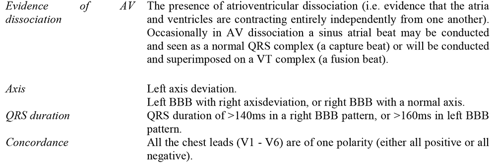
Table 4. ECG criteria suggestive of VT
You should then choose the most appropriate agent, guided by local availablility of drugs and what you believe to be the nature of the arrhythmia. Pharmacological treatments of the stable tachycardia include:
- Adenosine for supra-ventricular tachycardia (SVT). This drug is also useful if it is unclear whether the tachycardia is an SVT with a bundle branch block or VT. In SVT adenosine may convert the rhythm to sinus or slow it enough for the underlying rhythm to be established. It is have no effect on VT.
- Amiodarone for stable VT or SVT.
- Magnesium sulphate (2g IV) for torsades de pointe – a type of VT where the axis of depolarisation is continuously rotating.
- Seek expert help for irregular BCTs which may represent atrial flutter with a variable block or atrial fibrillation with rapid conduction due to an accessory conduction pathway.
This work by WFSA is licensed under a Creative Commons Attribution-NonCommercial-NoDerivitives 4.0 International License. To view this license, visit https://creativecommons.org/licenses/by-nc-nd/4.0/