Basic Sciences

QUESTIONS
Before continuing, try to answer the following questions. The answers can be found at the end of the article, together with an explanation. Please answer True or False:
- Regarding type 1 hypersensitivity reactions:
a. ‘Atopic individuals’ are at higher risk of developing type I hypersensitivity reactions
b. Require prior sensitisation to an antigen
c. Include anaphylactic reactions
d. Are mediated by IgG
e. Have one phase to the reaction - Anaphylactoid reactions:
a. Occur due to mast cell degranulation
b. Are the result of IgE cross-linking on mast cells
c. Are induced in all individuals exposed to anaphylactoid substances
d. Are differentiated from anaphylactic reactions by the absence of mast cell tryptase
e. May cause hypotension on induction of anaesthesia - Delayed hypersensitivity reactions are:
a. Also known as type IV hypersensitivity reactions
b. Occur within 2 hours of exposure to antigen
c. Comprise an infiltrate of T-helper cells and macrophages
d. May result in granuloma formation
e. Are most commonly seen in cutaneous reactions
Key Points
- Hypersensitivity describes the process of tissue damage secondary to an inflammatory reaction.
- Hypersensitivity reactions are broadly classed into type I-IV by the Gell and Coombes Classification.
- Anaphylaxis is an extreme form of a type I reaction that results due to widespread mast cell degranulation on interaction with IgE bound to antigen.
- The pathogenesis of a hypersensitivity reaction is often due to more than one class of hypersensitivity occurring concurrently.
INTRODUCTION
In the first immunology tutorial we covered the basic immunology that would help develop an understanding of where things can go wrong; these are the areas we will focus on in this tutorial. As anaesthetists we are only too aware of the risk posed by hypersensitivity in our patients and understandably are particularly fearful of anaphylactic reactions. Anaphylaxis is covered throughout anaesthetic training and the protocols for its management are familiar to all anaesthetists.
In this tutorial we will discuss the immunological principles underlying the four types of hypersensitivity reactions.
HYPERSENSITIVITY REACTIONS 1,2
During an immune reaction the release of inflammatory mediators results in increased vascular permeability and recruitment of inflammatory cells to cause local tissue inflammation. Hypersensitivity describes tissue damage secondary to an exaggerated inflammatory reaction. These reactions may be in response to an external antigenic challenges such as pollens in hay fever, an inappropriateresponse to a pathogen or even to an individuals’ own tissue.
Hypersensitivity reactions can be described by the Gell and Coombs classification. This was first put forwards as a classification in 1963 by immunologists Phillip Gell and Robin Coombs and still remains the most widely used classification for hypersensitivity. It classifies reactions depending on their timescale and aetiology producing four separate groups. Although the classes are apparently independent of each other, hypersensitivity reactions often involve more than one of the reactions at any one time.
TYPE I HYPERSENSITIVITY REACTIONS 1,2,3
Type I reactions are also known as immediate reactions and are seen in anaphylaxis, allergic asthma and eczema. In order to be at risk of type I hypersensitivity, an individual must have been sensitised to an antigen by an earlier exposure. First exposure would have produced an immunoglobulin E (IgE) specific to the antigen, the memory for that IgE is then held within the plasma cell repertoire. Hypersensitivity results when subsequent exposure to the antigen induces large scale production of IgE which binds to the Fcε receptors of mast cells. The interaction of the mast-cell bound IgE with their antigen induces degranulation and release of inflammatory mediators.
Mast cell granules contain histamine. When they degranulate, histamine release causes increased vascular permeability and vasodilatation which contributes to the immediate effects seen in type I hypersensitivity. Other factors released by degranulation are chemotactic factors, which are responsible for drawing in circulating eosinophils and basophils, and platelet activating factor which further contributes to the vascular changes seen in these reactions.
Although described as ‘immediate’ hypersensitivity, these reactions often have delayed component that occurs 4-6 hours later. This delayed reaction has two aetiologies; the synthesis of leukotrienes and prostaglandins which have similar vasoactive properties to histamine and the release of interleukin-4 from T helper 2 (Th2) cells which results in recruitment of further inflammatory cells. This second peak in the reaction is known as the late phase reaction and can last up to 24 hours following exposure. The use of corticosteroids in the immediate phase will reduce or abolish this secondary reaction.
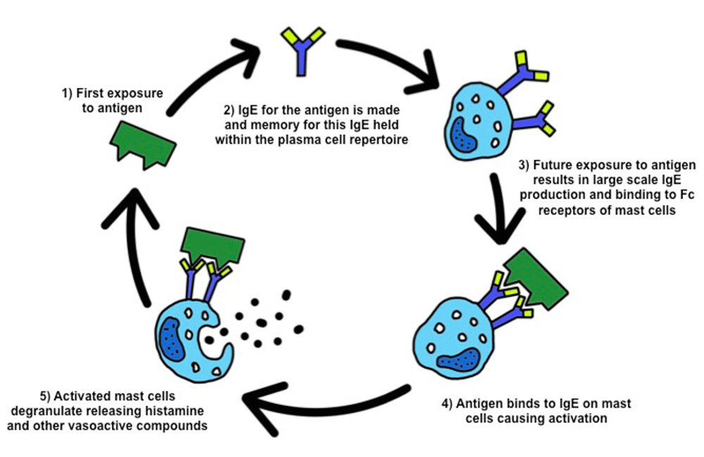
Figure 1: Diagrammatic representation of type I hypersensitivity reactions
Anaphylaxis 1,3,5
Most type I reactions occur locally as a result of IgE production to inhaled or ingested antigens as is seen with pollen or food allergies. Anaphylaxis is a dramatic example of a type I hypersensitivity reaction that results from the systemic administration of antigen.
The time course of anaphylaxis depends on the route of antigen delivery. If antigen is delivered systemically to a sensitised individual, for example through a bee sting or an intravenous drug, anaphylaxis ensues rapidly. When antigen is absorbed through the skin or gastro-intestinal tract, the onset of anaphylactic symptoms may be slower, for example with latex or peanut allergy.
Independent of the time course, the mechanism underlying the classical features seen in anaphylaxis is the same. The pathology is due to generalised mast cell degranulation and subsequent widespread histamine release following exposure to antigen in a sensitised individual. Histamine causes smooth muscle contraction, increased vascular permeability and vasodilatation which results in the classical signs seen in anaphylaxis such as urticarial rash, bronchospasm, facial swelling and cardiovascular collapse. The use of adrenaline can counteract the effects of histamine through alpha and beta agonism causing vasoconstriction and bronchodilation respectively. Adrenaline also contributes to a reduction in vascular permeability.
Anaphylactoid reactions 1,3
A common question that pops up in exams is the difference between anaphylactic and anaphylactoid reactions. Anaphylactoid reactions are anaphylaxis-like reactions that also result from mast cell degranulation and widespread histamine release. In these reactions the initial stimulation of mast cells does not occur through interaction of IgE with antigen. Instead mast cells are stimulated directly by certain substances. Examples of known anaphylactoid substances include dextran starches, fluorescein and radio-opaque contrast media. However, these reactions are not induced in all individuals exposed to these compounds.
Anaphylactoid reactions may be responsible for cases of severe cardiovascular collapse during anaesthesia; however the treatment of both anaphylactoid and anaphylactic reactions is the same and protocols for its management should be followed. The use of mast cell tryptase testing will only confirm mast cell degranulation and will not provide a differentiation between the two reactions. If anaphylactoid reaction is suspected, it is vital to seek expert immunological advice and exclude anaphylaxis.
Atopy1,4
Some individuals are more prone to allergic diseases than others due to an inherited pre-disposition to atopy. As discussed in the previous tutorial, Th2 cells are involved in the activation of B-cells to produce immunoglobulin and can encourage production of IgE. Atopic individuals have a shift in the balance of T-helper cells to a Th2 predominant response. This leads to an inherited tendency to produce IgE to common external antigens therefore these individuals are more likely to experience type I hypersensitivity reactions such as asthma, eczema and even anaphylaxis.
TYPE II HYPERSENSITIVITY REACTIONS 1,3
Type II hypersensitivity reactions are categorised as antibody mediated reactions. They occur when an antibody or immunoglobulin is produced in response to non-harmful antigens resulting in an unwanted immune response. As discussed in tutorial one immunoglobulins are vital for protection against foreign pathogens; They opsonise cells for destruction by leucocytes, whilst also recruiting further immune cells to result in an inflammatory reaction.
Type II hypersensitivity may occur when B-cells that produce immunoglobulin against native cells are not deleted during development. When this occurs, individuals are at risk of autoimmune disease including organ specific disease and immune haemolytic anaemias. An example of organ specific autoimmune disease due to type II hypersensitivity is Goodpasture’s syndrome. In this condition, antibodies are produced against type IV collagen found on the basement membrane of the lung and glomerulus. Binding of antibodies to collagen induces inflammation and subsequent damage to the basement membrane rendering it non-functional.
Other examples of type II mediated autoimmune diseases are autoimmune haemolytic anaemia where antibodies are specific for red blood cell antigens and myasthenia gravis where antibodies are produced against the acetylcholine receptor. Destruction of each target occurs in these conditions resulting in anaemia and neuromuscular weakness, respectively.
Some type II reactions occur in response to foreign antigens on transfused or transplanted cells. In these instances, although unwanted, the reaction is due a functioning immune system. The risk of transplant rejection and transfusion reactions is reduced by tissue matching the donor and recipient.
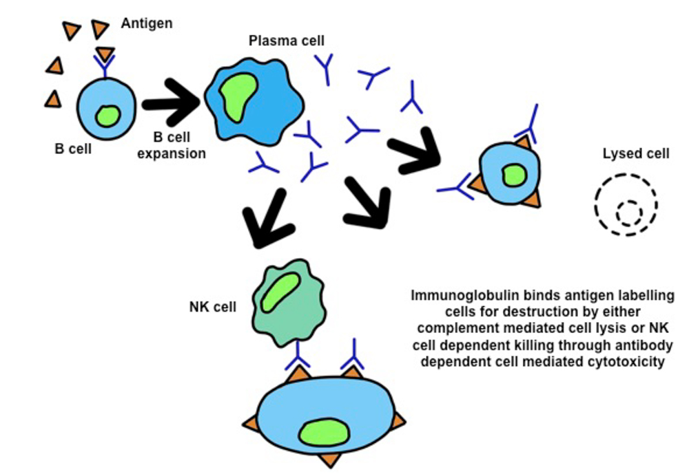
Figure 2: Type II Hypersensitivity reactions. The Presence of antigen stimulates specific B-cells. The B-cell undergoes clonal expansion and produces large amounts of immunoglobulin against the antigen. By binding antigen, the immunoglobulin labels the cell for destruction by NK cells or complement.
TYPE III HYPERSENSITIVITY REACTIONS 1, 3
Type III hypersensitivity reactions occur following the deposition of immune complexes, this results in complement activation and inflammation. For this reason, they are also known as immune complex mediated hypersensitivity reactions. In health, antigen:antibody complexes are maintained as soluble immune complexes in the blood by complement proteins C2 and C4. These immune complexes bind to complement receptors on red blood cells allowing their transport to the spleen where complexes are removed and destroyed. If the production of immune complexes is greater than the clearance, this is when pathology may arise.
When there is excessive production of immune complexes, precipitation may occur. This leads to complement activation, recruitment of immune cells and tissue damage through release of inflammatory mediators and free radicals. Areas affected by immune complex precipitation tend to be sites of filtration such as the glomeruli, synovium and epidermal basement membrane.
Some individuals have a higher predisposition to type III hypersensitivity; this can be due to inherited tendencies or preexisting disease processes. Inherited tendencies are due to C2 or C4 complement protein deficiency which causes increased precipitation due to reduced complex solubility. Systemic lupus erythematosus (SLE) is an example of a preexisting condition with higher risk of type III hypersensitivity. This may be due to reduced complement receptors on red blood cells leading to reduced rate clearance.
In some cases of type III hypersensitivity, the pathology is induced by high antigenic loads. This is seen in extrinsic allergic alveolitis or Farmer’s Lung, where inhaled organic dust particles produce large amounts of immune complexes in the lungs and causing inflammation of the alveoli. As mentioned, the glomeruli are common sites of type III hypersensitivity due to their function in filtration of plasma. There are a number of forms of immune complex mediated damage to the glomeruli but the most common is IgA nephropathy or Berger’s disease. This is the result of IgA complex deposition in the mesangium of the glomerulus. Immune complex deposition is also responsible for the renal damage seen in post-streptococcal glomerulonephritis and SLE.
Henoch-Schönlein purpura is a systemic illness characterised by vasculitic rash, painful joints, abdominal pain and occasionally glomerulonephritis. This condition also results from type III hypersensitivity and arises from the widespread deposition of IgA containing complexes.
Other disease states that are due to type III hypersensitivity include rheumatoid arthritis, where immune complexes deposit in the synovium of joints and dermatitis herpetiformis which causes a bullous skin reaction due to IgA complex deposition in the dermis.
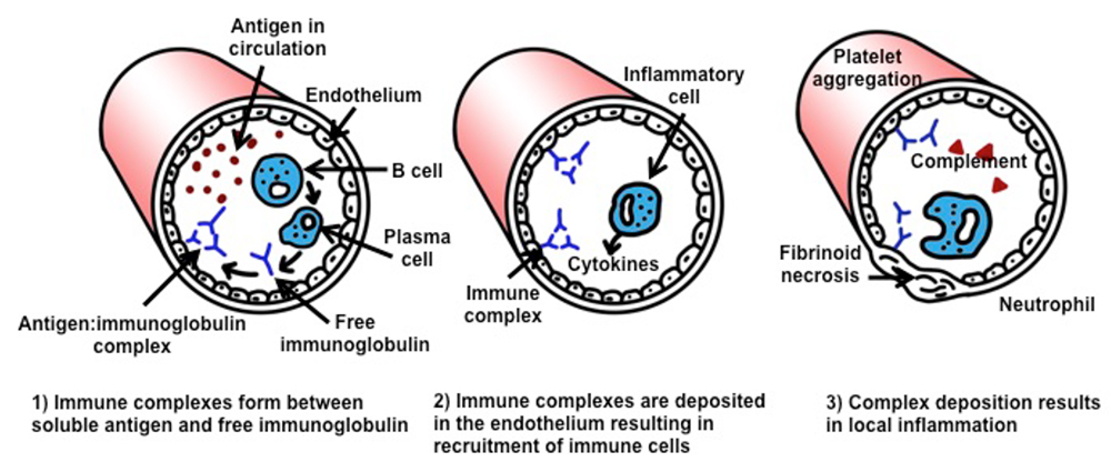
Figure 3: Pathogenesis of type III hypersensitivity.
TYPE IV HYPERSENSITIVITY REACTIONS 3,4,5
Type IV hypersensitivity reactions arise through the actions of stimulated antigen specific T-cells. These reactions may take up to twelve hours for features to develop and therefore are also known as delayed type hypersensitivity. Typically these reactions occur following contact of a sensitising antigen with the skin where the effects are seen as contact dermatitis. In these processes, antigen is taken up by local innate immune cells such as macrophages. These cells then act as antigen presenting cells and activate antigen specific CD4+ T-cells. T-cells activated in this manner tend to adopt a Th1 profile and migrate to the area of high antigenic load where they release inflammatory cytokines such as interferon-γ (IFN-γ), IL-1, IL-2 and IL-6. The result of the cytokines is local inflammation through an increase in vascular permeability and further immune cell migration and activation.
In a non-persistent antigenic challenge, the reaction tends to be maximal at 24-48 hours post exposure. If an antigenic challenge is prolonged, the ongoing inflammatory response can result in granuloma formation characterised by multinucleate giant cells produced by the fusion of macrophages. Granulomatous reactions can take weeks to resolve. Not all type IV reactions occur cutaneously. The same mechanisms seem to contribute to the development of type I diabetes and multiple sclerosis where Th1-cells mount an attack on β-cells of pancreas and myelin basic protein respectively. As a granulomatous disease, it is likely that type IV reactions are also involved in Crohn’s disease; however the trigger antigen is unknown.
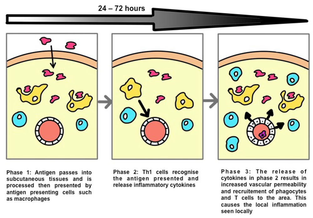
Figure 4: Pathology of cutanous type IV hypersensitivity.4
SUMMARY
An understanding of the immune processes underlying hypersensitivity will not only help with preparation for exams but will also help with everyday practice. These reactions may manifest as a minor type IV reaction from an adhesive dressing, or a severe anaphylactic reaction to systemically administered drugs.
The Gell and Coombs classification is a useful description of four differing aetiologies underlying the pathologies seen in hypersensitivity reactions. These processes are not always due to a disordered immune system and therefore some reactions require prevention with immunosuppressive agents – for example in autoimmune disease or prevention of transplant rejection. Although the classification describes the reactions as separate entities, in reality multiple aetiologies may be contributing to the clinical picture.
MCQ ANSWERS
- ..
a) True: Type 1 hypersensitivity reactions are more common in atopic individuals
b) True: Type 1 hypersensitivity reactions require prior sensitisation to an antigen
c) True: Anaphylaxis is a well described Type 1 hypersensitivity reaction
d) False: Type 1 hypersensitivity reactions are IgE mediated
e) False: Type 1 hypersensitivity reactions have an early and a delayed phase reaction - ..
a) True: Anaphylactoid reactions occur due to mast cell degranulation
b) False: Anaphylactoid reactions are independent of IgE
c) False: Anaphylactoid reactions only occur in some individuals exposed to certain triggers
d) False: Raised mast cell tryptase confirms mast cell degranulation and can occur in both anaphylactic and anaphylactoid reactions
e) True: Anaphylactoid reactions can be the cause of severe hypotension at the induction of anaesthesia - ..
a) True: Delayed hypersensitivity reactions are also known as type IV hypersensitivity reactions
b) False: Delayed hypersensitivity reactions are maximal at 24-48 hours post exposure
c) True: Delayed hypersensitivity reactions comprise an infiltrate of T-helper cells and macrophages
d) True: Delayed hypersensitivity reactions may result in granuloma formation
e) True: Delayed hypersensitivity reactions are most commonly seen in cutaneous reactions
REFERENCES
- Chapel H, Haeney M, Misbah S, Snowden N; Essentials of Clinical Immunology, 6th Ed. Oxford, Wiley- Blackwell, 2014
- Male, D; Immunology: An Illustrated Outline, 3rd Ed. London, Mosby-Wolfe, 1998.
- Todd I, Spickett G; Lecture Notes Immunology, 6th Ed. Oxford, Wiley-Blackwell, 2010
- Murphy K, Travers P, Walport M; Immunobiology, 7th Ed. New York, Garland Science, 2008.
- Male D, Brostoff J, Roth D, Roitt I; Immunology, 7th Ed. Philadelphia, Mosby Elsevier, 2006.
This work by WFSA is licensed under a Creative Commons Attribution-NonCommercial-NoDerivitives 4.0 International License. To view this license, visit https://creativecommons.org/licenses/by-nc-nd/4.0/