Paediatric Anaesthesia

INTRODUCTION
As intensivists in district general hospitals in the UK, every 2 to 3 months we are asked to assess and manage sick children within our hospital pending retrieval to a specialist paediatric unit seventy miles away. Intensivists who form part of the on-call ICU team have limited exposure to ICU during the daytime and generally encounter these patients at night. Dealing with sick children is one of the foremost challenges for an adult intensive care specialist and the following article serves as revision and reminders for colleagues who find themselves in similar situations in the middle of the night.
Stabilisation and retrieval of critically ill children is an exercise in team-work between you and your colleagues and the regional PICU (Paediatric Intensive Care Unit) team. It is vital you communicate with the PICU, seek advice for management of sick children at a very early stage and at regular intervals until the child is retrieved.
Detailed description, assessment and management decisions concerning individual disease processes is beyond the scope of this article. The points raised are from personal experience and constitute generic advice applicable to a wide variety of situations. Sedation and intubation forms an integral part of the overall management of the critically ill child as well as simultaneous attention to all aspects of resuscitation, including fluid management.
Sedation, intubation and ventilation may be indicated for children with airway pathology, airway compromise due to impaired consciousness or for respiratory failure. In addition, haemodynamic compromise that has failed to respond to fluid resuscitation is itself an indication for intubation and ventilation – up to 40% of a sick child’s cardiac output may be required to support its work of breathing.1 We should be aware that recent guidance suggests that initial fluid resuscitation in children with haemodynamic compromise, should occur rapidly – i.e. within the first 15 minutes from presentation. 1 Intubation and ventilation also aids safe insertion of invasive monitoring in critically ill coagulopathic children with severe sepsis.
When assessing a critically ill child, try to keep an open mind to the underlying diagnosis in each case, and beware the pitfall of assuming that the diagnosis is correct. For example, a child diagnosed with croup who deteriorates despite standard initial treatment may well have an alternative diagnosis such as epiglottitis (HiB vaccination failure is rare but not unknown) or bacterial tracheitis.
Avoid assuming that the working diagnosis is correct. View the child with a fresh pair of eyes and consider alternative diagnoses
LOCATION
Your initial assessment will usually take place in the emergency department or the paediatric ward (or paediatric high dependency unit (HDU) if your hospital has one). A consultant paediatrician will generally be present – if not they should be called. Following assessment of the child and discussion with the paediatric team it may be decided that the child should be sedated and intubated. In cases where this decision is unclear, it is useful to discuss with the regional paediatric retrieval centre – they may already be aware of this critically ill child.
The decision to move a critically ill child to an area of safety and familiarity (e.g. ICU or theatres) is one of the most difficult decisions to make. Advantages include a more controlled environment with familiar equipment, drugs and staff in which to proceed with sedation and intubation. Where there is airway compromise many would prefer to undertake an inhalational induction with a familiar anaesthesic machine in theatres. However, the potential for clinical deterioration in transit must be carefully considered, as deterioration may be rapid and cardio-respiratory collapse in the lift or corridor is a situation to be avoided at all costs.
The decision to intubate may be influenced by logistical issues. A child dependent on nasal CPAP (continuous positive airway pressure) who is deteriorating will need to be intubated at that site if the CPAP circuit cannot be moved with the patient. Also, consider that if the child is likely to be retrieved to a specialist centre (which will involve sedation and intubation) your threshold for undertaking intubation will be lowered.
If you decide that transfer is the preferred option, you must have available oxygen and all equipment needed for airway, breathing and circulatory support during transit. Make sure full monitoring (at least pulse oximetry and electrocardiogram) are used. If the child is comfortable, leave them as they are, but if they are distressed it is common practice to have a parent sitting on the bed, holding the child and delivering oxygen. Make sure that you feel in control of the situation and you are happy with the child’s position – you need to be able to see their face and have access to a limb to assess capillary refill time. If you have any doubts about the child’s condition, do not move them and if the situation changes during transfer return them to the original place of relative safety.
If you decide to move the child prior to intubation make sure that you feel in control during the transport and are adequately equipped to deal with any deterioration in the child’s condition
Finally, be aware that the paediatric team may not have cannulated a child with a diagnosis of an airway illness such as croup, tracheitis or epiglottitis. Intravenous induction is therefore not an option, making transfer inherently more hazardous. Remember that in extreme situations suxamethonium can be delivered via the intramuscular (IM), intralingual or intraosseous route (see Box 1)2 and intramuscular ketamine is effective in doses of 5-10mg.kg-1.
PERSONNEL
If intubation is planned, you should have the best available help with you. Where possible, induction and intubation of a sick child in a district hospital should be undertaken by two consultant anaesthetists/intensivists. It is advisable to ask your colleague on-call for the anaesthesia team to assist, no matter what their paediatric experience. The presence of an operating department practitioner (ODP) or an experienced ICU nurse is also invaluable.
Induction of anaesthesia and intubation of critically ill children is a two-consultant procedure. The most experienced anaesthetist should manage the airway.
At an early stage think whether there are other specialists who you may wish to be present, for example a consultant ENT surgeon in all cases of possible airway compromise.
Remember that for infants the paediatric consultant may have significant recent experience intubating babies in the neonatal ICU. Whilst it is important that trainees have the opportunity to be involved to gain experience, it is the authors’ recommendation that the responsibility for airway management and intubation should only be given to very senior trainees (senior registrar equivalent) and those with recent specialist experience of paediatric anaesthesia. However, this is almost always a job for a consultant – if intubation proves to be difficult and hypoxia results, valuable time is lost while personnel change positions. Consider whether re-allocating anaesthetists in theatre will give you more experienced help – for example a trainee may take over from a consultant in theatre and free the most senior member of the team to assist.
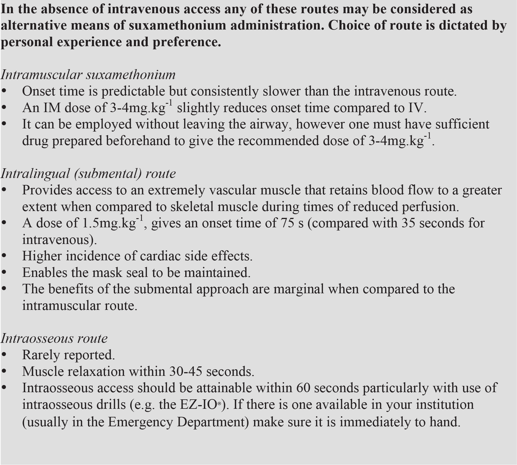
Box 1. Suxamethonium – alternatives to the intravenous route of administration2
Effective communication between members of the team is essential. Each member should be assigned specific roles, for example: an assistant to the doctor managing airway, a doctor responsible for administering drugs etc. Whilst it should be clear who is taking the lead role, remember to listen to the advice of others during the procedure, particularly if difficulties arise. It is difficult to remain objective under stressful circumstances and the situation may be worsened by persisting with an unsuccessful strategy. The goal is oxygenation, not intubation.
Ask a member of the nursing staff to document all events, procedures and drugs administered with accurate timing. This should be their only role.
Good communication between individuals and teams is essential – this includes listening to the advice offered by all staff present – particularly if the clinical situation becomes difficult.
It may be useful to nominate someone as a ‘runner’ – someone who is familiar with theatres and its equipment and who can retrieve items that are required unexpectedly.
PREPARATION/EQUIPMENT (see appendix 1 for checklist)
Logistics
Paediatric cots can be hard to negotiate from the head-end and it is often easiest to turn the child by 90° so that they are lying across the bed. The available space may be limited, so those without a specific role should be politely asked to vacate the room.
Introduce yourself to the parents and explain your role and what you plan to do. It is worth keeping one of them in the room until the child is asleep if it contributes to calm the child. Accepted practice is that the parents are escorted from the room just prior to induction of anaesthesia, intubation and subsequent stabilisation of the child. They should then be allowed to come back in as soon as is reasonably possible.
Drugs
Most PICUs will provide a ‘ready-calculator’ to provide a comprehensive list of all the drug doses and infusion mixes that you are likely to need. This should be immediately to hand.
In each case it is advisable to draw up the exact amount of each drug required in a 1ml syringe. Dilution of drugs can lead to confusion over doses and only needs to be considered if the child is very small (e.g. a premature neonate) or if 1ml syringes are unavailable.
Give Atropine a few minutes prior to induction
Anticholinergic agent
This is usually atropine at a dose of 20mcg.kg-1 (with a minimum dose of 100mcg). Hypoxia peri-intubation may be unavoidable with severe respiratory disease and a dose of atropine 2 to 3 minutes prior to induction will delay onset of the resultant bradycardic response to this. If ketamine is used, atropine reduces the hypersalivatory response to this drug. The authors would advocate use of atropine in this setting unless there is a specific contraindication.
Ketamine is a useful induction agent and analgesic, with a good safety profile in the critically ill.
Induction agent
The main alternatives are ketamine, thiopentone and propofol. The authors’ preference is ketamine, which has advantages of greater haemodynamic stability (due to sympathetic stimulation) and some inherent analgesic properties. Check the strength of ketamine you have been provided with (1, 10 or 100mg.ml-1).
Muscle relaxant (see Box 1)
Generally suxamethonium 2mg.kg-1 is the appropriate choice. Have the initial dose of a non-depolarising muscle relaxant (e.g. atracurium 0.5mg.kg-1) available to prolong muscle relaxation after the airway has been secured.
Fluid bolus
Have 20ml.kg-1 of crystalloid drawn up to treat haemodynamic instability. Consider giving this prior to induction of anaesthesia if there are signs of shock (e.g. prolonged capillary refill time). If greater than 40ml.kg-1 has already been administered consider using a colloid such as 4.5% albumin.
Ongoing sedation/anaesthesia
Ask your ICU to prepare infusions of sedative agents as guided by your regional specialist centre. In our region this is an infusion of fentanyl/vecuronium for younger children and morphine and midazolam infusions for other children. In the meantime additional increments of ketamine can be used (0.25-0.5mg/kg-1 lasts 10-15 minutes).
Emergency drugs
Prepare doses of 0.1ml.kg-1 of 1 in 10,000 epinephrine (adrenaline), for use in the event of cardiac arrest periinduction.
Airway equipment
The checklist in Box 3 shows suggested steps in preparation for intubation of a sick child. Further details concerning some items are contained within the text below.
Breathing circuit
Use the circuit you are most familiar with and an appropriately sized anaesthetic face mask – usually an Ayre’s Tpiece for children under 20kg, or a self-inflating (Ambu-) bag (Figure 1). Some paediatric units have paediatric versions of a Waters circuit – these are ideal for adults and larger children but are unwieldy for smaller children, as they require a ‘third hand’ to adjust the APL (adjustable pressure limiting) valve. The Ayre’s T-piece has the advantages of allowing application of CPAP and allowing you to assess and augment the child’s own respiratory effort. Self-inflating bags are almost universally available and are invaluable in the absence or failure of an oxygen source, when non-self-inflating systems are rendered redundant.
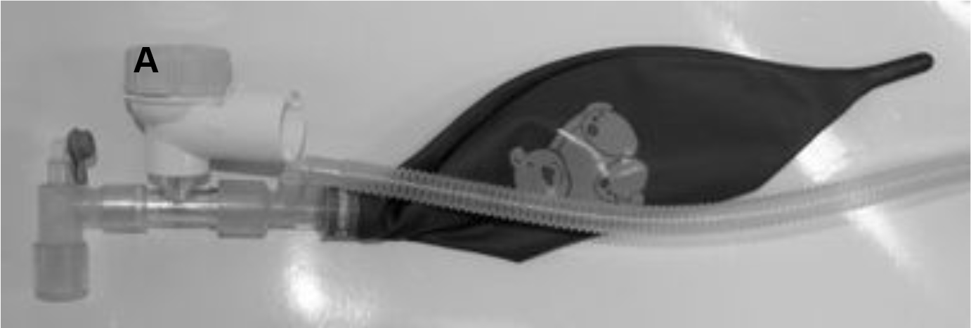
Figure 1. The paediatric Waters circuit is difficult to use for manual ventilation in small children since a ‘third hand’ is required to adjust the APL valve (A). It may have a role in provision of CPAP to children with spontaneous respiration
Suction
Have suction with a Yankauer attachment (preferably paediatric size) turned on and under the right-hand side of the child’s pillow. Have several suction catheters appropriate to the proposed endotracheal (ET) tube size (see Box 2) together with some pre-prepared aliquots of 1-2ml sterile saline to be used to loosen secretions.
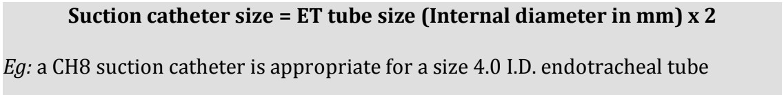
Box 2. Sizing endotracheal suction catheters
Children with a primary respiratory or airway problem (e.g. tracheitis or bronchiolitis) can prove very hard to ventilate after intubation is achieved. Lung compliance may be very poor due to lung consolidation (bronchiolitis) and may be so bad that you doubt the correct positioning of the ET tube. Capnography is very useful in these situations and should be used to confirm tracheal tube placement where available. A major contributor to poor respiratory compliance in airway and pulmonary diseases is the build up of thick secretions within the major airways. The ET tube may even become blocked during intubation. Airway lavage, chest physiotherapy and suction will resolve this problem, but it may take several attempts – a senior ICU nurse will be invaluable in doing this effectively.
In smaller children, inflation of the stomach during resuscitation/assisted ventilation may hinder ventilation once intubated. Drainage of a gas filled stomach via a naso-gastric (NG) tube may improve ventilation markedly. In contrast to historical practice, it is often advisable to use a cuffed endotracheal (ET) tube in critically ill children, in order to achieve adequate ventilation.
Have suction equipment available – children with severe respiratory or airway disease will often be very difficult to ventilate even when intubation is achieved.
Endotracheal tubes
Prepare the appropriate size with one size larger and smaller (two sizes smaller in airway obstruction). Tubes below about size 5.0 may be difficult to pass between the vocal cords due to their flexibility and we recommend that they are premounted on a shapable stylet.
Use a stylet to ease passage of smaller ET tubes through the cords.
A large proportion of difficulties encountered after intubation are due to the ET tube being over- or under-inserted (i.e. too long or too short). We recommend cutting the ET tube after intubation and confirmation of appropriate positioning of the tip of the tube by chest X-ray. This is particularly important in facial burns where significant swelling can be expected. Remember:
Length of ET tube at gums/incisors = Age/2 + 12cm
For smaller children many PICU prefer nasal intubation; however we recommend initial oral intubation in all children unless there are no cardiorespiratory concerns (e.g. a child with an isolated intracranial lesion). The tube can be changed to a nasal tube when they are stable or you can defer this procedure to the retrieval team. A recent study of over 2000 children under 5 years of age undergoing anaesthesia for elective surgery, suggests that the rate of post extubation stridor is no greater when a cuffed compared to an uncuffed tracheal tube is used.3 Where available use of a cuffed tracheal tube in sick children will provide a more reliably sealed airway, allow more effective ventilation. The need tube changes (when the leak around the tube is excessive) is also reduced.3
Once placed the ETT must be secured – this is best achieved using two ‘trouser leg’ lengths of elastoplast (see Figure 3). Be aware that small tubes are easily kinked and obstructed during or after this process.
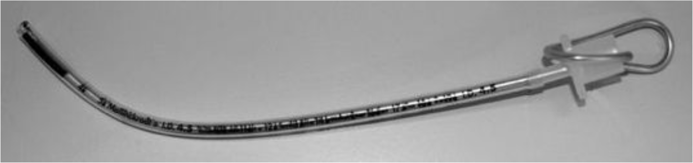
Figure 2. Size 4.5mm ET tube with stylet
Monitoring
Use capnography to confirm correct ET tube position within the trachea.
Use all of the monitoring that is available and if time allows bring a transport monitor from ICU. The importance of capnography has been emphasised above. If you are unfamiliar with the equipment find out who is and ask for their assistance. Set the pulse oximeter to an audible tone mode, which you can hear. Alternatively ask a staff member to call out the child’s saturations every 10 seconds during intubation. Use your ‘runner’ to fetch any vital equipment or drugs that you need from ICU or theatres.
If intubation is difficult remember that the objective is oxygenation, not intubation and revert to bag-mask ventilation
POST-INTUBATION
When you have confirmed that the ET is in the trachea, check the length to the child’s lips and fix the tube securely using elastoplast ‘trousers’ as shown in Figure 3.
Check that your intravenous access is secure and plan further access if this will be needed for ongoing fluid resuscitation and sedation. Useful sites include the long saphenous vein just anterior to the medial malleolus at the ankle and the femoral vein for multiple channel access. This latter site should only be attempted in small children if you have some experience of the technique in this age-group. Similarly arterial access may be useful, via the radial or femoral artery, but unsuccessful attempts at various sites will make you unpopular with the retrieval team when they arrive!
Ongoing haemodynamic instability after adequate fluid resuscitation may necessitate inotropic support. Dobutamine or dilute epinephrine may be delivered peripherally with caution, but dopamine or epinephrine via a central venous catheter is the preferred treatment. It is sensible to obtain the advice of the retrieval team prior to starting these drugs.
Other aspects of the child’s care to consider are:
- Update the retrieval team and ask for their expected time of arrival.
- Blood sugar – check this periodically and treat hypoglycaemia.
- Place a urinary catheter and naso-gastric tube if needed.
- Keep the parents informed and document this. They should be in the room as much as they wish to be and as much as is logistically possible.
- Document all events accurately with timings – use the notes of the staff member you nominated to do this.
- Prescribe all of the drugs you have given and all of those that need to be continued (e.g. antibiotics and infusions). Check that appropriate antibiotics have been administered.
- Request any further investigations that are possible now the child is asleep, such as throat swab for viral PCR etc.
- Chest X-ray to confirm ET tube position, NG tube position and lung pathology.
- Debrief all nursing and medical staff involved in the child’s care, getting their feedback on the events.
Remember to use the expertise of the paediatric team – they have far greater familiarity with tasks such as prescription of maintenance fluids.
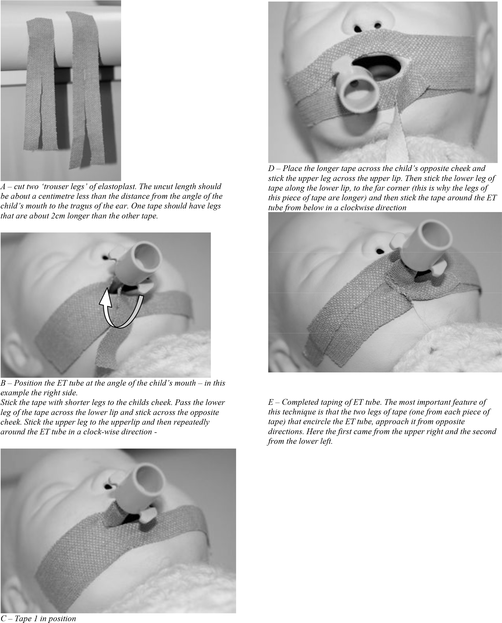
Figure 3. Securing an endotracheal tube with tape.
SUMMARY
Those considering a career in adult ICU in a district hospital, without specialist PICU facilities, will be called upon to assist in the management of critically ill children. It is highly recommended to spend some time training in a PICU – the experience gained can be useful many years later. Many PICUs run refresher courses (e.g. the Splat course, http://www.bmsc.co.uk/courses/SPLAT.htm) or it is usually possible to arrange to spend some time on your regional unit shadowing the permanent staff. The PICU in our region participates in ‘Road-show’ days where recent cases are discussed. It is beneficial to attend local morbidity and mortality meetings and debrief sessions organised by paediatric and emergency departments. It is also worth discussing cases with junior staff and nursing staff, particularly if the outcome for the child had been bad. Take time to discuss your view of events with a colleague.
REFERENCES
- Brierly J, Carcillo JA, Choong K et al. Clinical practice parameters for haemodynamic support of pediatric and neonatal septic shock: 2007 update from the American College of Critical Care Medicine. Crit Care Med 2009; 37: 666-88.
- Walker RWM, Sutton RS. Which port in a storm? Use of suxamethonium without intravenous access for severe laryngospasm (editorial). Anaesthesia 2007; 62: 757-59.
- Weiss M, Dullenkopf A, Fischer JE, Keller C, Gerber AC and the European Paediatric Endtracheal Intubation Study Group. Prospective randomized controlled multi-centre trial of cuffed or uncuffed endotracheal tubes in small children. BJA 2009; 103: 867-73.
ACKNOWLEDGMENTS
Many thanks to Daniel Lutman, Consultant for Children’s Acute Transport Service, and Pete Ford, Consultant in Intensive Care Medicine, for reading and improving this tutorial.
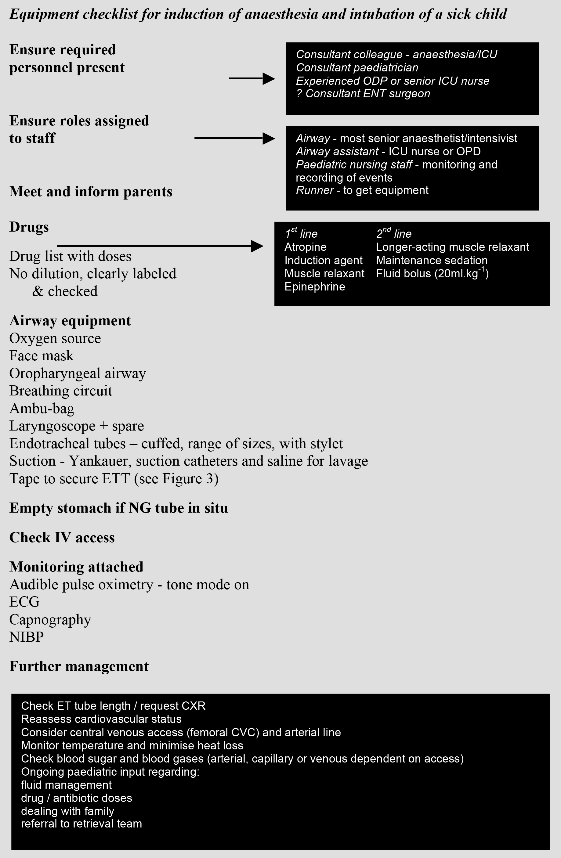
Appendix 1: Preparation checklist for induction of anaesthesia and intubation of a sick child
This work by WFSA is licensed under a Creative Commons Attribution-NonCommercial-NoDerivitives 4.0 International License. To view this license, visit https://creativecommons.org/licenses/by-nc-nd/4.0/