Obstetrics Anaesthesia

KEY POINTS
Platelet Disorders
- Gestational thrombocytopaenia is the commonest cause of mild thrombocytopaenia during pregnancy.
- Patients with platelet disorders undergoing neuraxial procedures are at increased risk of epidural haematoma potentially resulting in cord compression and neurological dysfunction.
Congenital Coagulopathies
- Von Willebrand disease, haemophilia A (factor VIII deficiency), and haemophilia B (factor IX deficiency) are the commonest congenital coagulopathies seen in pregnant women.
- Monitoring factor levels and determining the presence of a bleeding history are important in assessing risk of haemorrhage and complications from neuraxial procedures.
- Factor levels should be monitored throughout pregnancy and the postpartum period, and replaced to achieve levels greater than 50% of normal.
- Patients may be at risk of primary and secondary postpartum haemorrhage due to the rapid decline in coagulation factors in the peripartum period.
INTRODUCTION
Pregnant women with platelet and/or coagulation disorders are more susceptible to bleeding complications, especially during the antepartum and postpartum periods and should be managed by a multidisciplinary team that includes haematologists, obstetricians, and anaesthetists. An individualised delivery plan should be established during early pregnancy with special consideration to appropriateness of neuraxial analgesia or anaesthesia and mitigation and management of potential postpartum haemorrhage (PPH).
PLATELET DISORDERS
Thrombocytopaenia
Thrombocytopaenia (a platelet count < 150 × 109/L) is common during pregnancy, occurring in approximately 7% to 10% of pregnant women.1 The majority of thrombocytopaenic cases during pregnancy are mild (130-150 × 109/L), have normal platelet production and function, and are not associated with adverse maternal, foetal, or neonatal outcomes. Gestational thrombocytopaenia is the commonest cause, occurring in 75% of women presenting with a low platelet count.1
Other conditions associated with thrombocytopaenia in pregnancy include the following:
- Preeclampsia (~ 20%) and haemolysis, elevated liver enzymes, and low platelets (HELLP) syndrome ( 0.17% to 0.85%)
- Immune thrombocytopaenic purpura (ITP) (~ 4%)
- Thrombotic thrombocytopaenic purpura (TTP) and haemolytic uraemic syndrome
- Disseminated intravascular coagulation (DIC), acute fatty liver of pregnancy, infections, and malignancies (~ 1%-2%).
Gestational Thrombocytopaenia
Gestational thrombocytopaenia occurs in 7% to 10% of pregnancies. The platelet count is approximately 75 to 100 × 109/L and platelet function is normal. It occurs due to the dilutional effect of increased plasma volume, and a hormonal effect leading to increased consumption of platelets in the placental circulation. It is not associated with clinical signs of bleeding and does not affect the foetus.
Preeclampsia and HELLP Syndrome
Preeclampsia is a multisystem disorder. Diagnostic criteria of preeclampsia are as follows:
- New-onset hypertension (systolic blood pressure ≥ 140 mm Hg and/or diastolic blood pressure ≥ 90 mm Hg) after 20 weeks of gestation and proteinuria ≥ 300 mg in a 24-hour urine collection, or protein:creatinine ratio ≥ 0.3.2
- In the absence of proteinuria, preeclampsia is diagnosed as hypertension associated with one or more severe features, such as new-onset thrombocytopaenia, impaired liver function, renal insufficiency, pulmonary oedema, or visual or cerebral disturbances.2 Of note, proteinuria is no longer a prerequisite for diagnosis.
The exact pathophysiology is unknown, but it is thought to be due to abnormal placentation, defective invasion of spiral arteries of the uterus by cytotrophoblastic cells, and alteration in the vascular response to nitric oxide leading to increased vascular tone.3 Thrombocytopaenia occurs in up to 50% of women with preeclampsia and its severity increases with the severity of preeclampsia.1
HELLP syndrome is a serious complication of pregnancy. It is characterised by haemolysis, elevated liver enzymes, and low platelet count. The incidence is 0.17% to 0.85% of all pregnancies; risk of recurrence in a subsequent pregnancy is approximately 19% to 27%.4 Microangiopathic red cell and platelet destruction results in haemolysis and low platelet count. Obstruction of hepatic sinusoids with fibrin strands are responsible for elevated liver enzymes. HELLP syndrome is associated with high maternal mortality (eg pulmonary oedema, cerebral haemorrhage, renal failure, hepatic rupture, and placental abruption), and foetal mortality (eg preterm delivery and low birth weight).4 DIC occurs in > 80% of severe cases of preeclampsia or HELLP.1
Immune Thrombocytopaenic Purpura
ITP accounts for approximately 4% of pregnant patients presenting with thrombocytopaenia. It is characterised by a platelet count < 100 × 109/L. Patients may be diagnosed prior to pregnancy due to increased bleeding tendencies, or it may present as an incidental finding with a first trimester complete blood count. Formation of IgG autoantibodies against platelet surface antigens leads to increased destruction of platelets; platelet function is not affected (and may be increased). Significant spontaneous maternal haemorrhagic complications are not common unless the platelet count is < 20 × 109/L.5
Symptomatic patients, those with severe thrombocytopaenia (platelet count < 30 × 109/L), or patients about to undergo invasive procedures should be treated. Treatment options include corticosteroids and intravenous immunoglobulin, which may take several days to increase the platelet count.5 A recommended aim is to maintain the platelet count > 50 × 109/L for vaginal or caesarean delivery.5 If delivery is imminent or in the setting of acute haemorrhage a platelet transfusion may be required.5 Splenectomy (ideally during the mid–second trimester to reduce risks to the foetus) may be required for patients with severe thrombocytopaenia not responding to medical therapy. IgG autoantibodies cross the placenta and may lead to significant foetal and neonatal thrombocytopaenia.
TTP and Haemolytic Uraemic Syndrome
TTP is a life-threatening condition with a mortality rate of 90% in untreated cases.6 Pregnancy in these patients should be managed at a tertiary centre. TTP presents with microangiopathic haemolytic anaemia, thrombocytopaenia (in the range of 10-30 × 109/L), neurological symptoms, fever, and renal abnormalities.1,6 It is caused by deficiency of von Willebrand factor (vWF) cleaving protein ADAMTS13.6 In the absence of this protein, platelet aggregates are not cleared and cause microvascular obstruction in vital organs.
Haemolytic uraemic syndrome has a similar pathophysiology to TTP but predominantly causes renal impairment.
Following are guidelines on managing TTP during pregnancy1,6:
- The ADAMTS13 (vWF cleaving protease) level is monitored throughout pregnancy and the postpartum period.
- Plasma exchange should be considered.
- Early delivery may be required; this does not guarantee reversal of symptoms.
- Treatment with corticosteroids and monoclonal antibodies is reserved for patients unresponsive to plasma exchange.
- Platelet transfusion is contraindicated, unless there is active haemorrhage.
- Thromboprophylaxis should be started (if the platelet count is > 50 × 109/L).
- Red cell transfusion and folic acid supplements are recommended during active haemolysis.
Disseminated Intravascular Coagulation
DIC is a consumptive coagulopathy which can be triggered by obstetric and nonobstetric aetiologies and is associated with a significant maternal morbidity and mortality (up to 25%).7
It is characterised by tissue factor–mediated activation of the coagulation cascade and fibrinolytic mechanisms which result in intravascular fibrin deposition, depletion of platelets and coagulation factors, and increased fibrin degradation products. DIC clinically presents with a wide range of bleeding abnormalities. Precipitants in pregnancy include placental abruption, significant PPH, HELLP syndrome, amniotic fluid embolism, sepsis, and intrauterine foetal death. Principles of management include early diagnosis, treatment of the underlying cause, and management of coagulopathy with administration of blood products (as indicated by haematological and coagulation parameters).
GUIDELINES FOR NEURAXIAL ANAESTHESIA IN PATIENTS WITH THROMBOCYTOPAENIA8
- Safety of neuraxial techniques are guided by expert opinion; risk of vertebral canal haematoma should be balanced with risks of general anaesthesia in the obstetric patient.
- The absolute platelet count, as well as anticipated platelet function (the aetiology of thrombocytopaenia and presence of a bleeding history) in addition to the rate of decline of platelets are important considerations (eg, a patient with severe preeclampsia where the platelet count had fallen from 150 to 90 × 109/L over 6 hours is likely more at risk than a patient with ITP with a stable platelet count of 70 × 109/L).
- A stable platelet count > 100 × 109/L is considered safe for neuraxial analgesia or anaesthesia.
- In preeclampsia, platelet counts may change rapidly; a recent (within 6 hours) count should be confirmed prior to a neuraxial procedure (including epidural catheter removal); a coagulation screen should be performed if the platelet count is < 100 × 109/L, or if the patient has deranged liver function tests.
- In gestational thrombocytopaenia and ITP, platelet function is normal; neuraxial blocks may be considered on a risk-benefit basis between counts of 50 and 100 × 109/L. There are several suggested ways to further reduce risk in these patients:
- A ‘‘low-impact’’ procedure should be performed by the most experienced anaesthetist available.
- Preprocedure ultrasound is potentially useful, if the provider is skilled in this.
- A single-shot spinal likely involves less risk than an epidural (smaller needle, often less traumatic insertion, no catheter left in situ; movement of the catheter may cause trauma of blood vessels or there may be bleeding on removal).
- If neuraxial procedures are undertaken, increased neuromonitoring until full recovery is essential; using a low-dose local anaesthetic solution for labour analgesia aids early identification of an unexpected motor block and concern about haematoma formation (this would facilitate an early magnetic resonance imaging scan, and if necessary timely neurosurgical intervention).
- With platelet count < 50 × 109/L the risks likely outweigh the benefit of neuraxial procedures regardless of aetiology.
COMMON CONGENITAL COAGULOPATHIES
Von Willebrand Disease
Description
This is the most common inherited coagulation disorder (incidence 1:100).9 It is inherited as an autosomal dominant trait and characterised by defective platelet adhesion and aggregation.
vWF is a glycoprotein synthesised by megakaryocytes and endothelial cells. It plays a role in primary haemostasis (adhesion of platelets to the vessel wall at the site of vascular injury) and secondary haemostasis (by acting as a carrier for factor VIII and stabilising it). The disease is classified into 3 main types (see Table 1).
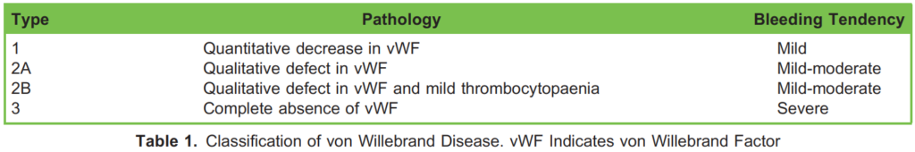
Pregnancy is usually associated with a rise in vWF, therefore levels in patients with type 1 von Willebrand disease (vWd) improve during pregnancy. Seventy-five percent of vWd cases are type 1. Patients diagnosed with types 2 and 3 are at higher risk of bleeding and should be managed by a multidisciplinary team at a tertiary centre. Factor levels should be monitored during pregnancy and labour. The vWF and factor VIII levels should be maintained above 0.5 IU/mL.9,10
Anaesthetic Considerations for vWD
Evidence in regard to neuraxial blockade comes from multiple case series in patients with type 1 disease.9 If vWF levels are normal in patients with type 1 disease, neuraxial labour analgesia can be safely offered.11 In patients with type 2 disease, neuraxial analgesia may be considered if the vWF and factor VIII levels are normal.11
Neuraxial anaesthesia should be avoided in patients with type 3 disease. The vWF levels need to be considered at the time of epidural catheter removal. Patients should be closely monitored for secondary PPH due to the rapid fall in vWF levels postdelivery. In cases of active bleeding, treatments include tranexamic acid, desmopressin (1-deamino-8-D-arginine vasopressin), replacement of specific factors, fresh frozen plasma, and cryoprecipitate. Desmopressin is contraindicated in type 2B disease as it can exacerbate thrombocytopaenia. Desmopressin is associated with hyponatraemia; if administered, plasma sodium levels should be checked regularly in conjunction with considered fluid administration.
Haemophilia
Description
Haemophilia A and B are X-linked disorders that affect mainly males; females are heterozygous carriers and have a baseline of 50% normal factor levels. Females are usually asymptomatic.
Haemophilia A is a deficiency of factor VIII. Factor VIII levels normally rise during pregnancy, so patients with haemophilia A usually have relatively normal factor levels and do not suffer from bleeding complications. In heterozygous carriers, factor VIII levels are commonly as low as 50% of normal but can be lower due to inactivation of one of the X chromosomes (due to lyonisation). Laboratory tests show low factor VIII activity, prolonged activated partial thromboplastin time and normal prothrombin time. Antenatally, factor VIII level should be maintained at > 50% of normal levels by factor VIII concentrate or cryoprecipitate.
Haemophilia B (Christmas disease) is a deficiency of factor IX. Laboratory tests show low factor IX activity but normal factor VIII activity and prolonged activated partial thromboplastin time. Replacement with factor IX concentrate or fresh frozen plasma may be necessary to maintain factor IX levels around 50% of normal levels. Factor IX levels do not increase in pregnancy, therefore there is a greater risk of bleeding complications with haemophilia B compared with haemophilia A peripartum.
In acute haemorrhage, recombinant factor VIIa has successfully been used in the treatment of haemophilia.
Factor XI Deficiency (Haemophilia C)
This is a rare disorder with autosomal dominant and recessive inheritance (however heterozygotes can have bleeding tendencies), predominantly seen in Ashkenazi Jews. There is a poor correlation between factor levels and risk of bleeding. The incidence of spontaneous bleeding is low, even with very low factor levels, but there is an increased risk of bleeding following surgery or trauma.11 It causes a prolonged activated partial thromboplastin time. Pregnant women with this disorder are at increased risk of antepartum and PPH so replacement with factor XI concentrate should be considered. Fresh frozen plasma can also be administered.
Anaesthetic Considerations for Patients With Haemophilia A or B
A detailed history of severity of symptoms and treatment should be taken. The platelet count, coagulation profile and factor respective VIII or IX levels should be obtained. Factor levels > 0.5 IU/mL (> 50% activity) are considered safe for neuraxial procedrues.9 Factor VIII should be replaced aiming for normal levels prior to caesarean delivery. Desmopressin may help increase factor VIII levels, and factor IX concentrate can be administered to increase factor IX levels.11
Risks of neuraxial procedures should be considered versus the risk of general anaesthesia, and patients should be counselled in the antenatal period regarding options for labour analgesia and surgical anaesthesia. This also include alternative options for labour analgesia such as opioid-based patient-controlled intravenous analgesia or nitrous oxide (50%):oxygen (50%). Deep intramuscular injections should be avoided due to risk of developing intramuscular haematomas. Use of nonsteroidal antiinflammatory drugs should be avoided because of their deleterious effect on platelet function.
Factor VIII levels fall rapidly after delivery and patients are at increased risk of secondary PPH, so factor levels should be maintained within the normal range for 3 days post–vaginal delivery and 5 days post–instrumental vaginal delivery or caesarean delivery.11
SUMMARY
Management of platelet disorders and congenital coagulopathies during pregnancy can be challenging. It needs early involvement of a multi-disciplinary team to assess the severity of illness and planning the management during pregnancy, labour and post-partum period. Central neuraxial blocks and options for labour analgesia should be carefully considered depending on platelets levels, coagulation profile and coagulation factors levels during labour.
REFERENCES
1. Myers B. Diagnosis and management of maternal thrombocytopenia in pregnancy. Br J Haematol. 2012;158(1):3-15.
2. American College of Obstetricians and Gynecologists; Task Force on Hypertension in Pregnancy. Hypertension in pregnancy: report of the American College of Obstetricians and Gynecologists’ Task Force on Hypertension in Pregnancy. Obstet Gynecol. 2013;122(5):1122-1131.
3. Uzan J, Carbonnel M, Piconne O, et al. Pre-eclampsia: pathophysiology, diagnosis, and management. Vasc Health Risk Manag. 2011;7:467-474.
4. Aloizos S, Seretis C, Liakos N, et al. HELLP syndrome: understanding and management of a pregnancy-specific disease. J Obstet Gynaecol. 2013;33(4):331-337.
5. Ciabanu A, Colibaba S, Cimpoca B, et al. Thrombocytopenia in pregnancy. Maedica (Buchar). 2016;11(1):55-60.
6. Scully M, Hunt BJ, Benjamin S, et al. Guidelines on the diagnosis and management of thrombotic thrombocytopenic purpura and other thrombotic microangiopathies. Br J Haematol. 2012;158(3):323 335.
7. Cunningham FG, Nelson DB. Disseminated intravascular coagulation syndromes in obstetrics. Obstet Gynecol. 2015;126(5):999-1011.
8. Association of Anaesthetists of Great Britain & Ireland The Obstetric Anaesthetists’ Association Regional Anaesthesia UK. Regional anaesthesia and patients with abnormalities of coagulation. Anaesthesia. 2013;68(9):966-972.
9. Katz D, Beilin Y. Disorders of coagulation in pregnancy. Br J Anaesth. 2015 Dec;115 (suppl 2):ii75-88.
10. Reynen e, james p. von willebrand disease and Pregnancy: A Review of Evidence and Expert Opinion. Semin Thromb Hemost. 2016;42(7):717-723.
11. Pavord S, Rayment R, Madan B, et al on behalf of the Royal College of Obstetricians and Gynaecologists. Management of inherited bleeding disorders in pregnancy. Green-top Guideline No. 71. BJOG 2017; 124:e193-e263.
This work by WFSA is licensed under a Creative Commons Attribution-NonCommercial-NoDerivitives 4.0 International License. To view this license, visit https://creativecommons.org/licenses/by-nc-nd/4.0/